This website uses cookies to ensure you get the best experience on our website.


- Table of Contents
Optimizing antigen retrieval in immunohistochemistry (IHC) is critical for unmasking epitopes that become concealed during formalin fixation and paraffin embedding. This step enhances the binding efficiency of primary...
Antigen retrieval is essential in immunohistochemistry (IHC) due to protein cross-linking caused by formalin fixation, which masks antigenic sites and hinders antibody binding. Formaldehyde fixation usually generates methylene bridges which cross-link proteins and therefore mask the epitope of interest. It is essential to unmask the antibody...
The purpose of using a secondary antibody is to amplify the primary antibody and antigen interactions. Before choosing a secondary antibody for your experiment, ask yourself the following 3 questions to help you choose the appropriate one:
The secondary antibody should be raised against the host in which the primary antibody was raised. For example, if the primary antibody was raised in rabbit, you will need an anti-rabbit secondary antibody raised in a species other than rabbit.
The secondary antibody has to be directed specifically against the isotype of the primary antibody. For example, the polyclonal primary antibodies are generally of the IgG isotype and thus the corresponding (species specific) secondary antibody needs be an anti-IgG antibody. The monoclonal
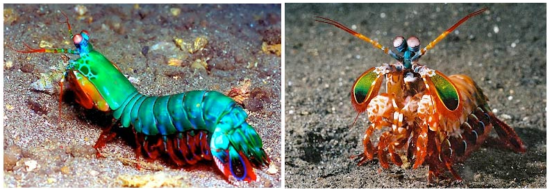
Did you know...The mantis shrimp has SIXTEEN color-receptive cones which allow it to see a MASSIVE spectrum of color that human brains aren't even capable of processing (we only have 3 types of cones). Good thing the mantis shrimp will never get to design multicolor flow cytometry antibody panels... and good thing Boster has 6 helpful tips for humans to make multicolor panels right here! Click here...
The Fluorescence Minus One (FMO controls) are staining controls that contain all the antibodies of a panel minus 1 of them. It measures the spillover of all those other fluorophores in the channel of the missing antibody, and is used to identify and gate cells in the context of data spread due to the multiple fluorophores. It is a stronger negative control than the regular unstained control as it takes into account how the other stains in the panel impact the channel that is left out.
There are 3 situations when an FMO control is highly recommended! Click here for more FACS controls information.
With the addition of every new color, the chances of non-specific spillover increases in practically

This month, we highlight several research advancements in tissue engineering and regenerative medicine, including the novel role of a transiently activated epicardial progenitor cell to enhance cardiac repair, inhibition of AMPK-mediated fibro-adipogenic progenitors apoptosis for muscle regeneration, and inhibition of PTGFR and its downstream pathway for proliferative retinal disease management.
In addition, recent discoveries in cancer diagnosis and treatment are presented. Different approaches to enhance cancer cells' response to immunotherapy, such as combining PCIF1 inhibition with anti-PD-1 treatment, offer promising results for tumor growth suppression.
Scroll down to learn more about these research studies!
Authors: Xia, Y., Duca, S., Perder, B., Dündar, F., Zumbo, P., Qiu, M., Yao, J…
Journal: Nature Communications
The epicardium, a mesothelial cell tissue that encompasses vertebrate hearts, supports heart regeneration after injury through paracrine effects and as a source of multipotent progenitors. However, the progenitor state in the adult epicardium has yet to be defined. Through single-cell RNA-sequencing of isolated epicardial cells...
Cited Boster Product(s): Anti-Zebrafish Mef2 Antibody (DZ01398-1)

Authors: Zhao, Y., Lei, Y., Ning, H., Zhang, Y., Chen, G., Wang, C., Wan, Q…
Journal: EMBO Molecular Medicine
The pathological retinal angiogenesis often causes blindness. Current anti-angiogenic therapy for proliferative retinopathy targets the vascular endothelial growth factor (VEGF), but many patients do not radically benefit from this therapy. Herein, we report that circulating prostaglandin (PG) F2α metabolites were increased in type...
Cited Boster Product(s): Mouse CXCL1/Gro Alpha ELISA Kit PicoKine® (EK0723)

Authors: Wang, L., Wu, L., Zhu, Z., Zhang, Q., Li, W., Gonzalez, G. M…
Journal: The EMBO Journal
Adenosine N6-methylation (m6A) and N6,2′-O-dimethylation (m6Am) are regulatory modifications of eukaryotic mRNAs. m6Am formation is catalyzed by the methyl transferase phosphorylated CTD-interacting factor 1 (PCIF1); however, the pathophysiological functions of this RNA modification and PCIF1 in cancers are unclear...
Cited Boster Product(s): Mouse TGF-Beta 3 ELISA Kit PicoKine® (EK1104)

Authors: Liu, X., Zhao, L., Gao, Y., Chen, Y., Tian, Q., Son, J. S., Chae, S. A…
Journal: Journal of Cachexia, Sarcopenia and Muscle
Background: Following muscle injury, fibro-adipogenic progenitors (FAPs) are rapidly activated and undergo apoptosis at the resolution stage, which is required for proper muscle regeneration. When excessive FAPs remain, it contributes to fibrotic and fatty infiltration, impairing muscle recovery. Mechanisms controlling FAP apoptosis...
Cited Boster Product(s): Mouse TGF Beta 1 ELISA Kit PicoKine® (EK0515)

Authors: Wang, Z., Qin, X., Hu, D., Huang, J., Guo, E., Xiao, R., Li, W., Sun, C...
Journal: Cell Reports
Ovarian cancer (OC) remains a clinical challenge for its difficulty in early diagnosis and insensitivity to treatments. Gut microbiota modulate multiple carcinoma progression through immunoregulation. The relationship between OC and gut microbiota has not been fully characterized. We find that the feces of patients with OC demonstrate...
Cited Boster Product(s): Mouse IFN Gamma / IFNG / Interferon Gamma ELISA Kit PicoKine® (EK0375)

Authors: Zhao, D., Yuan, H., Fang, Y., Gao, J., Li, H., Li, M., Cong, H…
Journal: International Journal of Biological Sciences
Evidence has indicated that lysine methyltransferase 2B (KMT2B), a major H3K4 tri-methyltransferase (H3K4me3), contributes to the development of various cancers; however, its role in cervical cancer (CC) is unclear. In this study, increased KMT2B expression was observed in human CC specimens and significantly associated...
Cited Boster Product(s): Human EGF ELISA Kit PicoKine® (EK0325)

Authors: Isildar, B., Ozkan, S., Ercin, M., Oktayoglu, S. G., Oncul, M…
Journal: Inflammation and Regeneration
Type 1 diabetes (T1D) is a T-cell-mediated autoimmune disease characterized by the irreversible destruction of insulin-producing β-cells in pancreatic islets. Helper and cytotoxic T-cells and cytokine production, which is impaired by this process, take a synergetic role in β-cell destruction, and hyperglycemia develops due to insulin...
Cited Boster Product(s): Human IFN Gamma ELISA Kit PicoKine® (EK0373); Human IL-17/IL-17A ELISA Kit PicoKine® (EK0430); Human IL-4 ELISA Kit PicoKine® (EK0404); Human IL-10 ELISA Kit PicoKine® (EK0416)

Authors: Guo, X., Du, L., Ma, N., Zhang, P., Wang, Y., Han, Y., Huang, X…
Journal: Journal of Translational Medicine
Background: Radiation-induced lung injury (RILI) often occurs during clinical chest radiotherapy and acute irradiation from accidental nuclear leakage. This study explored the role of monophosphoryl lipid A (MPLA) in RILI. Materials and Methods: The entire thoracic cavity of C57BL/6N mice was irradiated at 20 Gy with or without...
Cited Boster Product(s): Mouse

Counting cells before the staining procedure and the analysis/live sort is not up for debate! Cell numbers affect the staining quality, the FACS instrument reading, as well as the efficacy of any downstream assay, in case the cells are live sorted. Furthermore, cells are steadily lost during the staining procedure and the recovery of most sorters is a little above 50% − it is therefore imperative that enough cells are available at the start of the experiment to compensate for the inevitable losses. There are various methods available for counting cells:
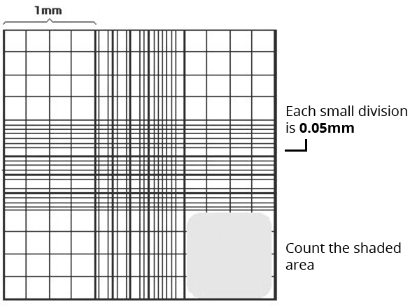
Hemocytometers are manual counting chambers and were first developed for blood cell counts. It consists of a thick glass slide onto which a gridded chamber is affixed. The four corner squares of the grid measure 1 mm x 1 mm and each square is further subdivided into 0.05 mm x 0.05 mm squares. A glass coverslip, made to specifications of the hemocytometer, can be placed 0.1 mm above the marked grid, thereby creating a space of known volume.
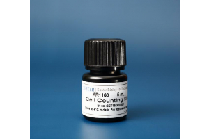
The cells are counted using a 4x or 10x objective lens over an area of 1 sq mm and the counting is repeated for a total of four such areas and the result is averaged. The formula for counting cells for each area is (average number of cells * 10 4 * dilution factor). To count the viable cells, use Trypan Blue exclusion method. Save some time and trouble with Boster’s ready-to-use Trypan Blue Assay Kit AR1175, see the kit below!.
Although the easiest method and least expensive, it is also labor intensive and allows for inaccuracies of the human eye. The image below depicts a typical hemocytometer counting grid.
They perform on the same principle as the hemocytometers but with the added advantages of speed and precision. Automated cell counter models have been developed by man...
Nitrocellulose membranes are one of the top matrices used in protein blotting in Western Blotting, offering significant advantages over simple filter paper in terms of protein retention and signal clarity. They have high protein-binding capacity, strong affinity for proteins of varying protein size, compatibility with a variety of protein detection methods, and the ability to immobilize proteins, glycoproteins, or nucleic acids. This variety of detection methods include chemiluminescence, chromogenic, and fluorescence. It is proven to produce excellent signal-to-noise results when used for amino acid analysis, protein sequencing and western, northern, and Southern blotting.
Western blotting (also called Protein Immunoblotting) is an analytical technique used to detect specific proteins in the given sample. A thorough understanding of the western blot principle is crucial to successfully applying this method and accurately interpreting the results, while proper western...

Are you preparing for a FACS experiment? Here’s a 10 point checklist to help you prepare for your FACS sorting experiment: