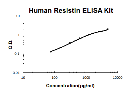
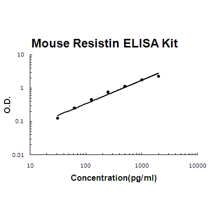
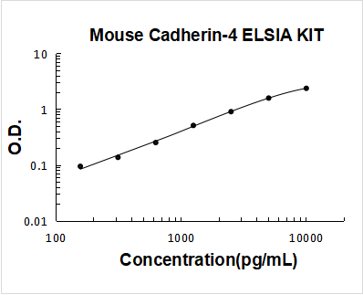
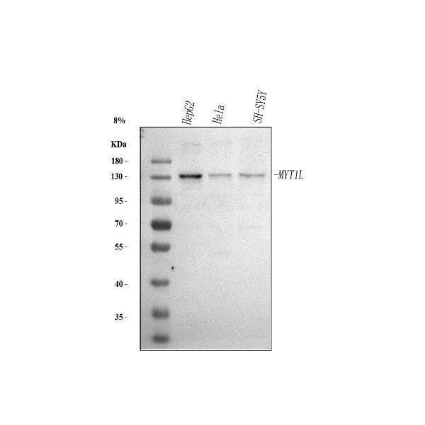
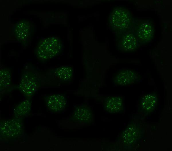
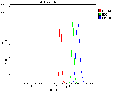
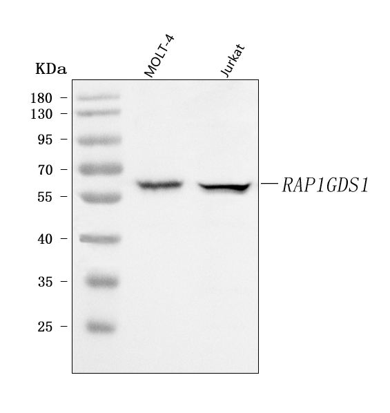
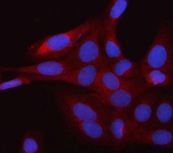
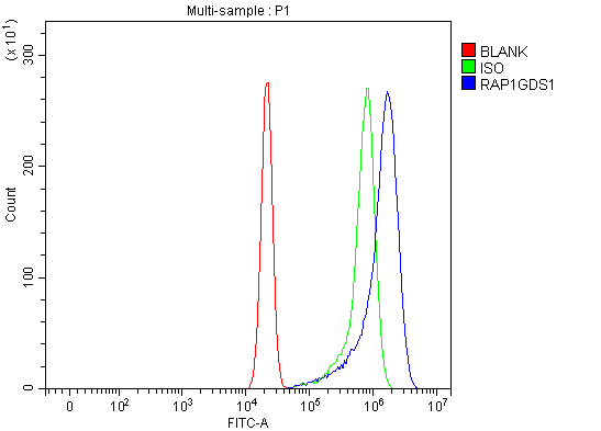
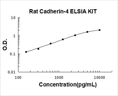
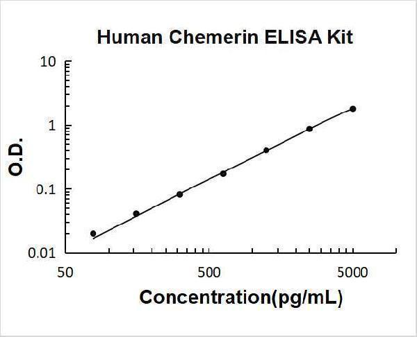
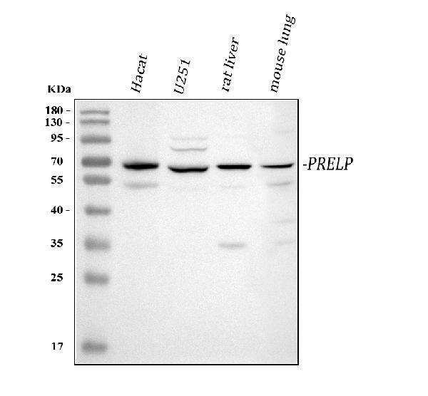
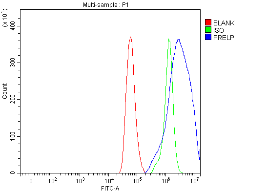
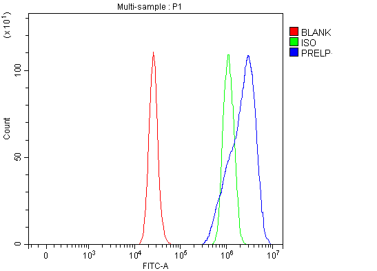
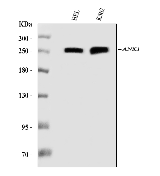
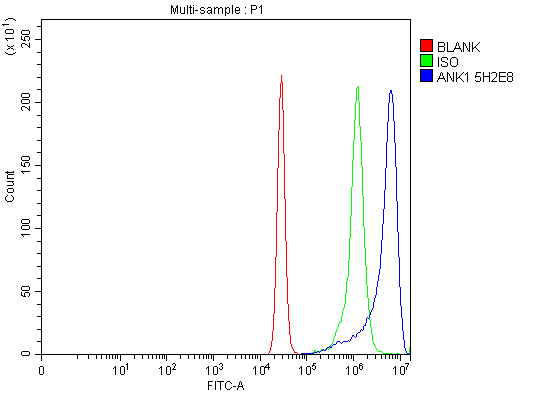
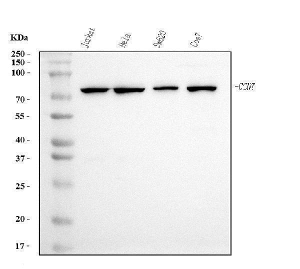
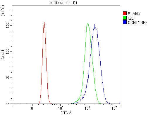
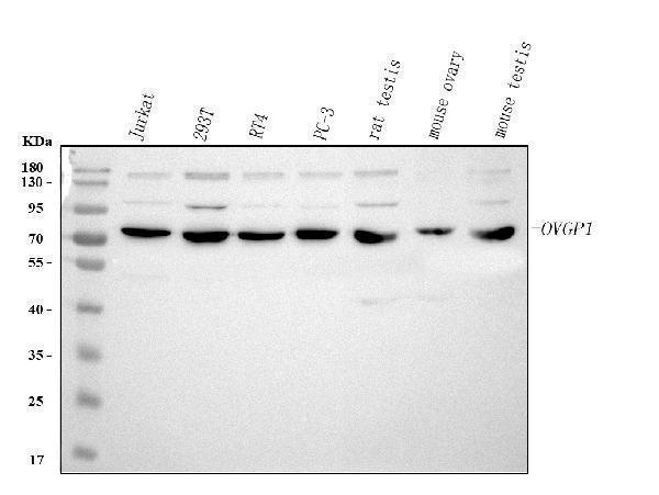
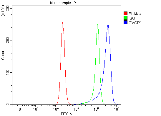
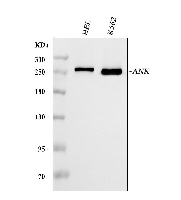
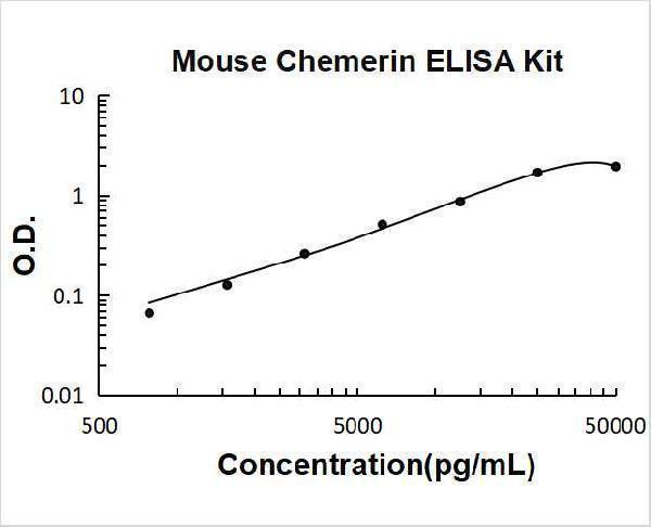
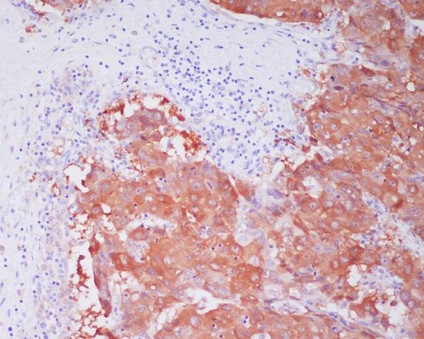
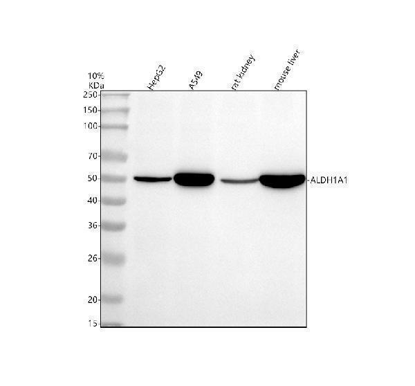
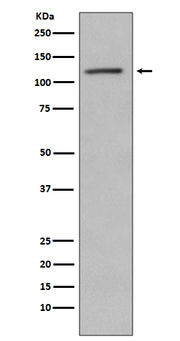
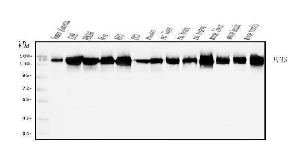
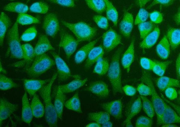
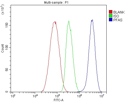
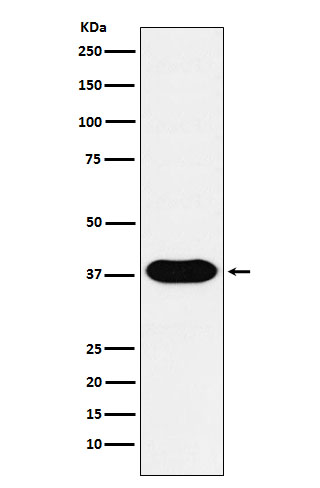
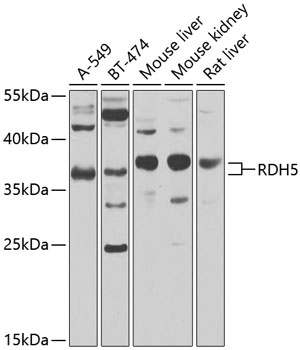
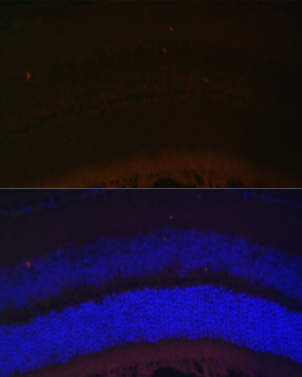
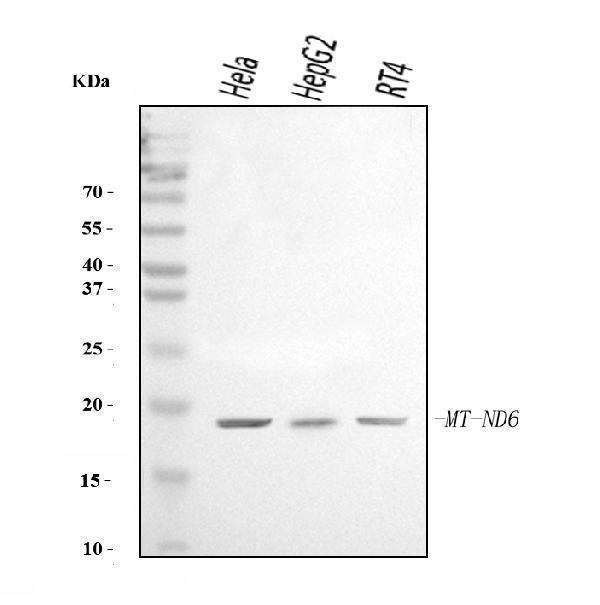
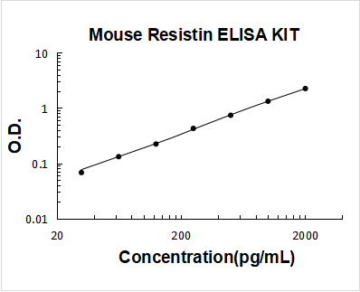
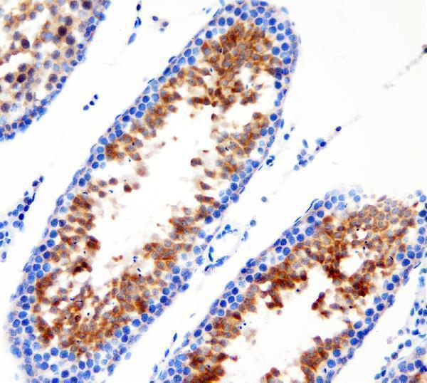
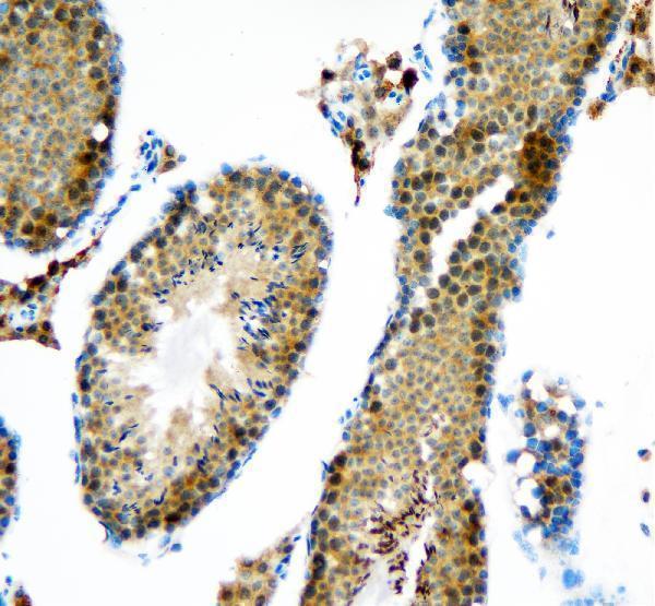
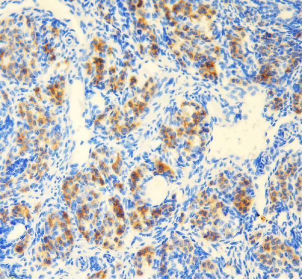
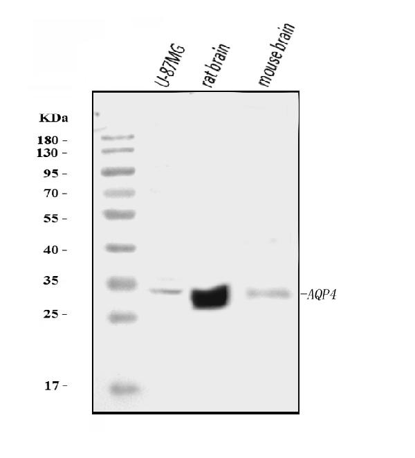
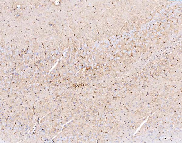
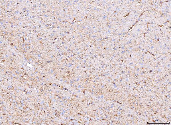
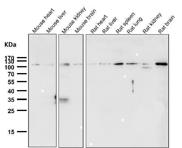
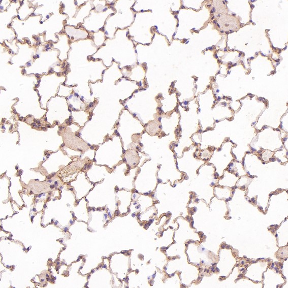
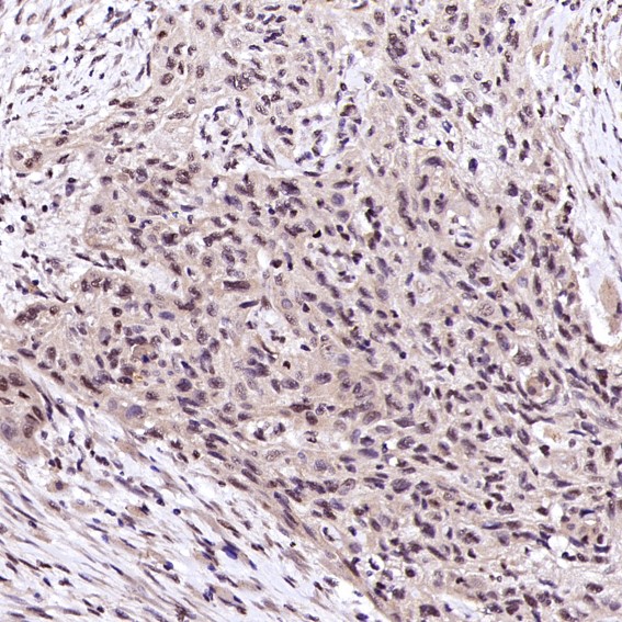
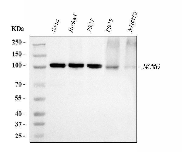
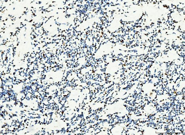
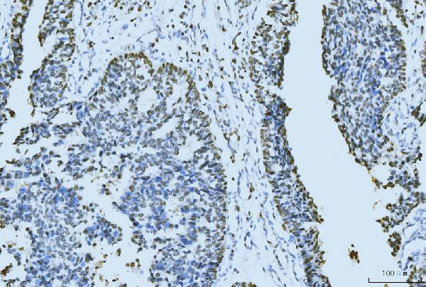
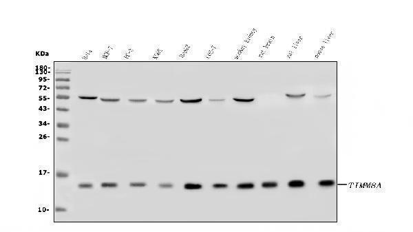
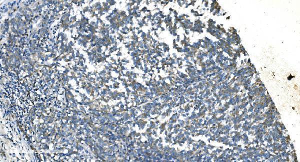
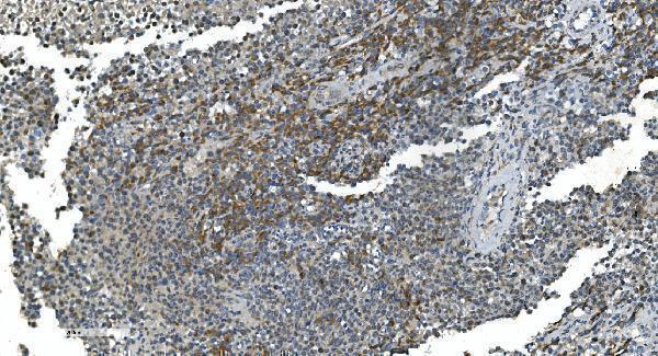
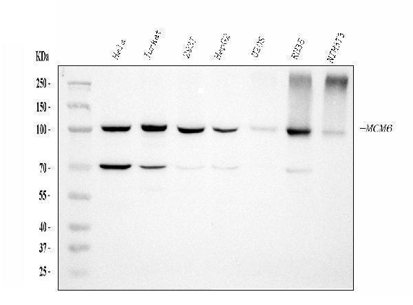
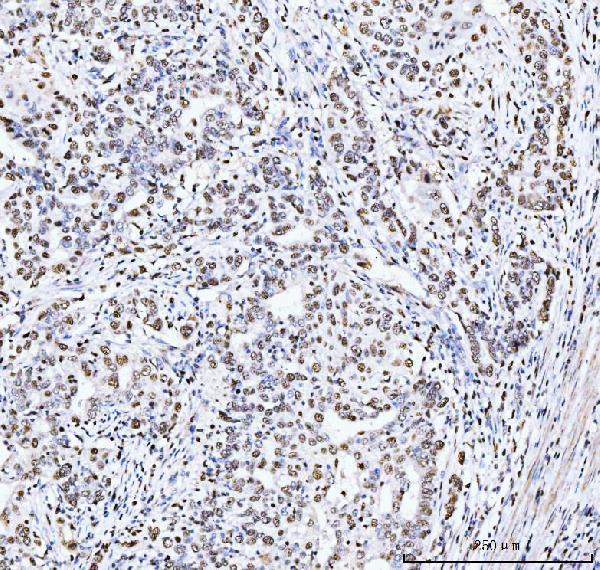
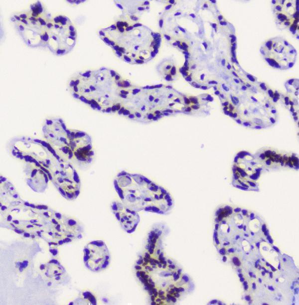
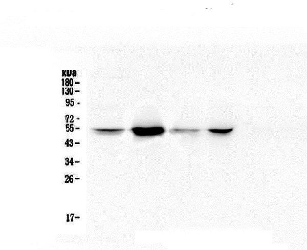
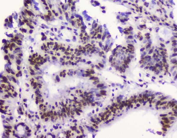
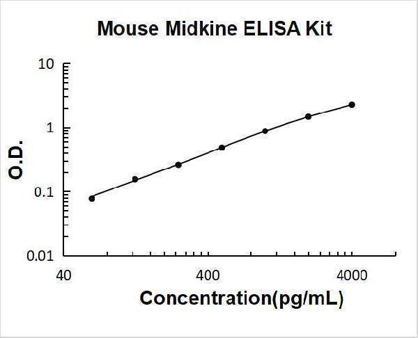
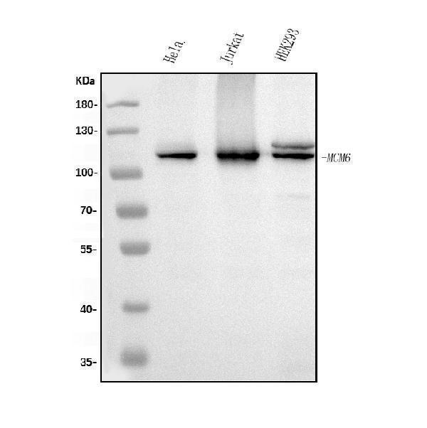
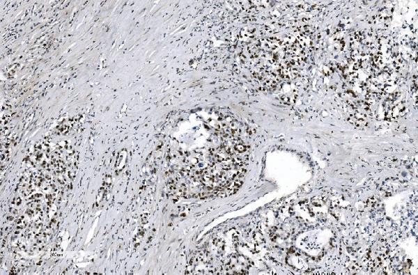
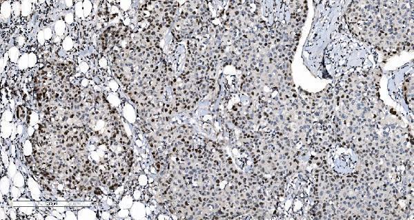
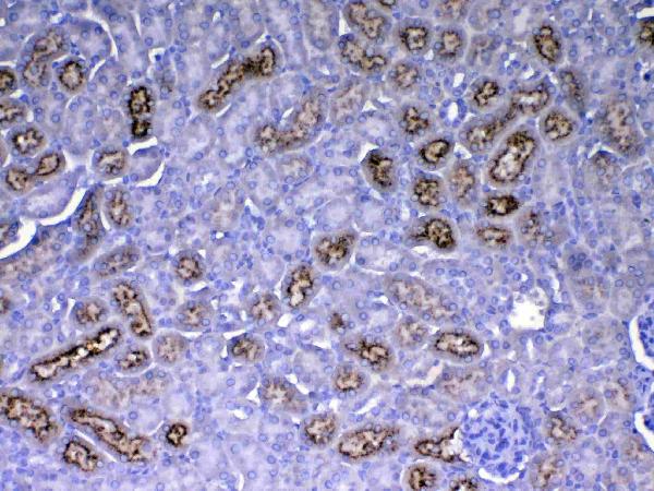
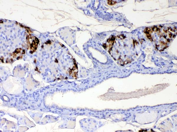
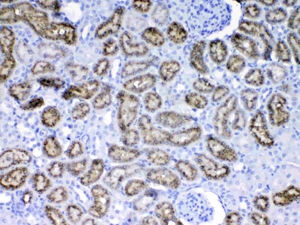
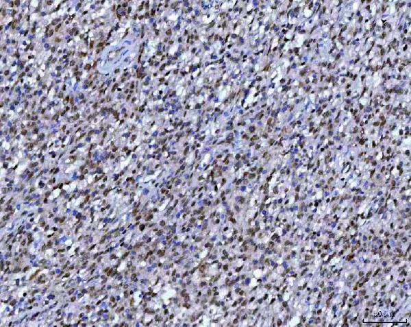
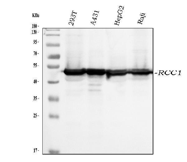
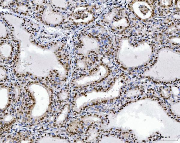
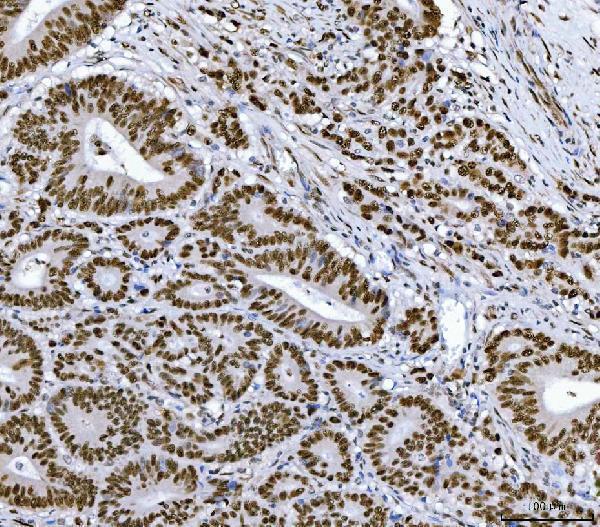
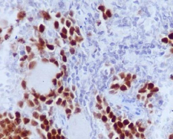
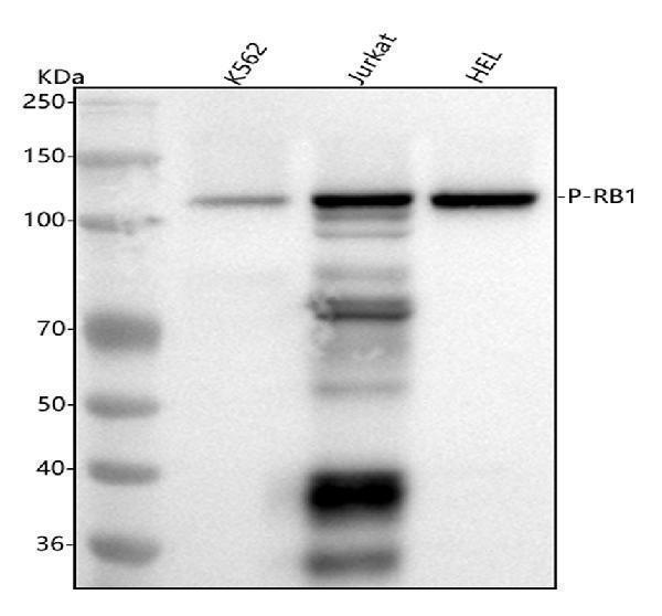
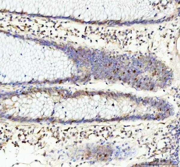
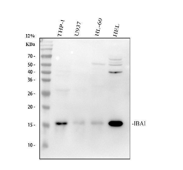
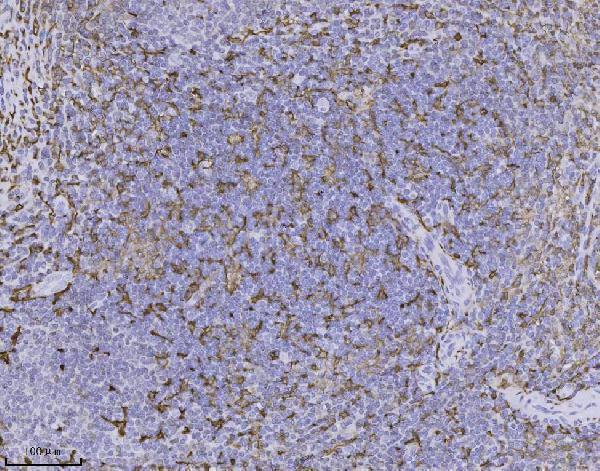
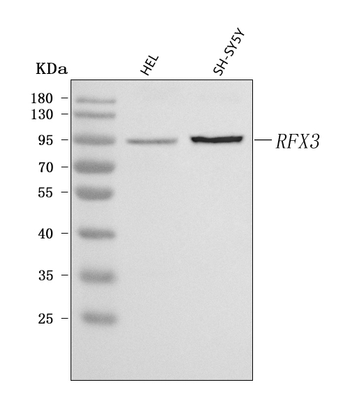
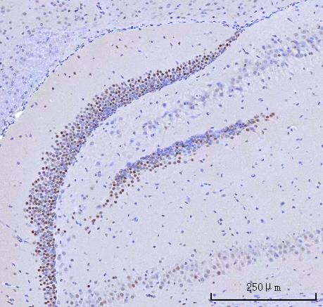
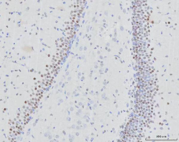
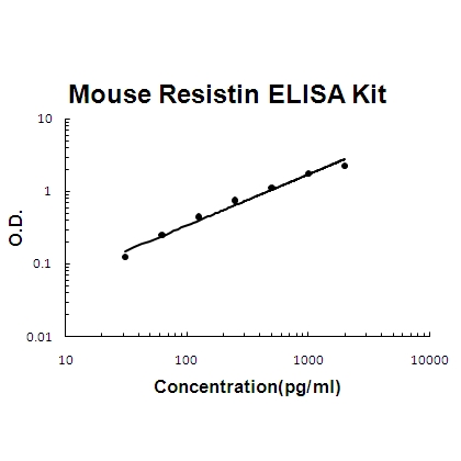
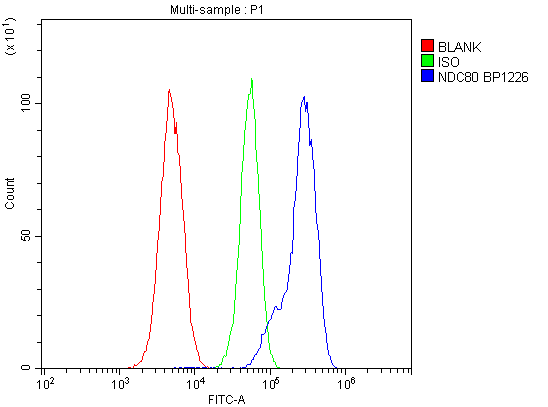
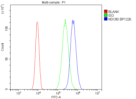
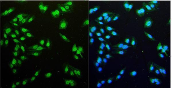
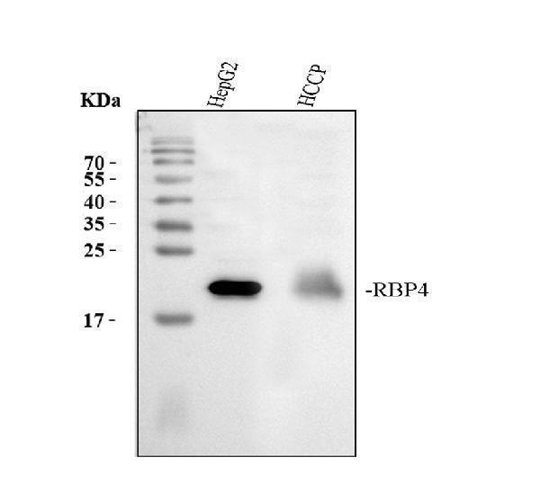
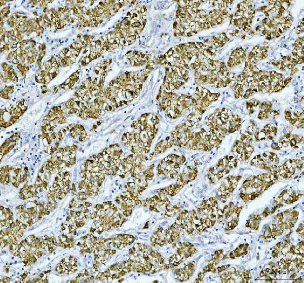
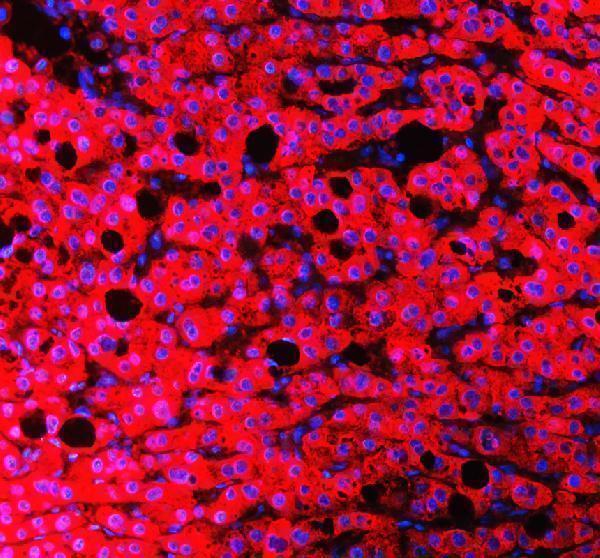
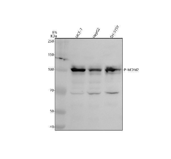
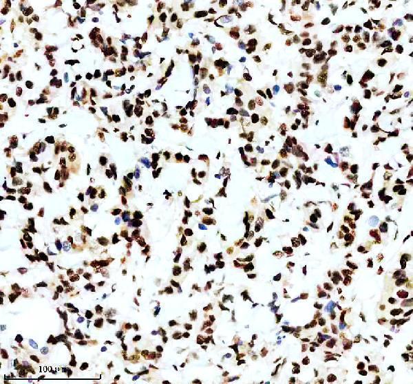
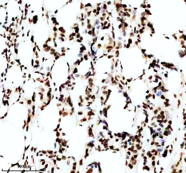
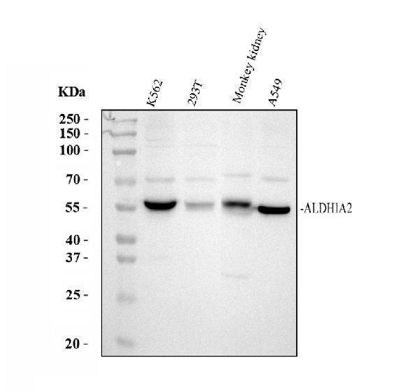
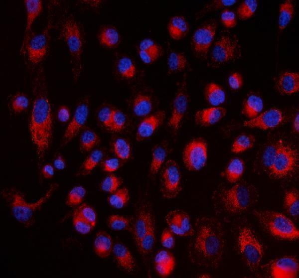
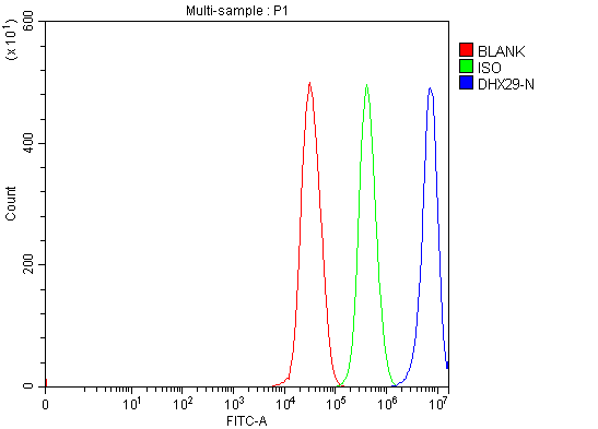
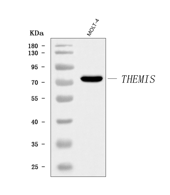
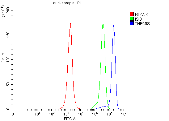
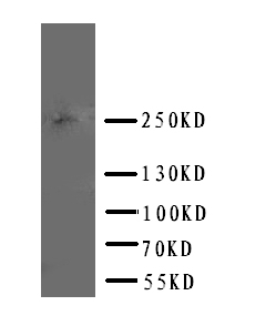
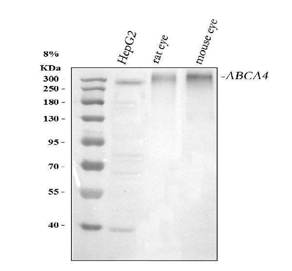
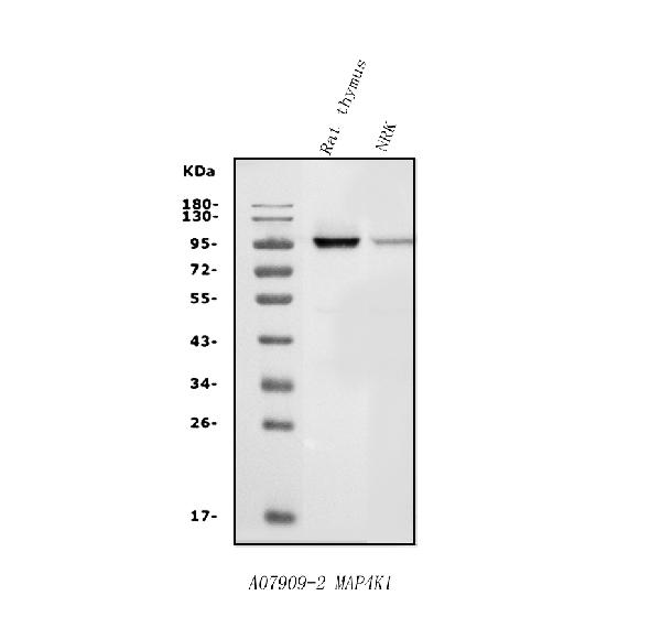
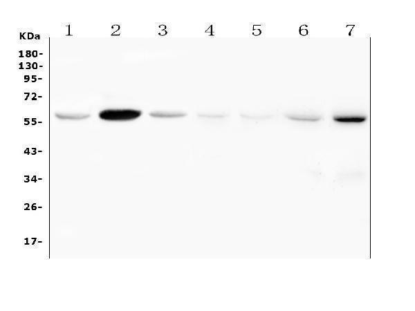
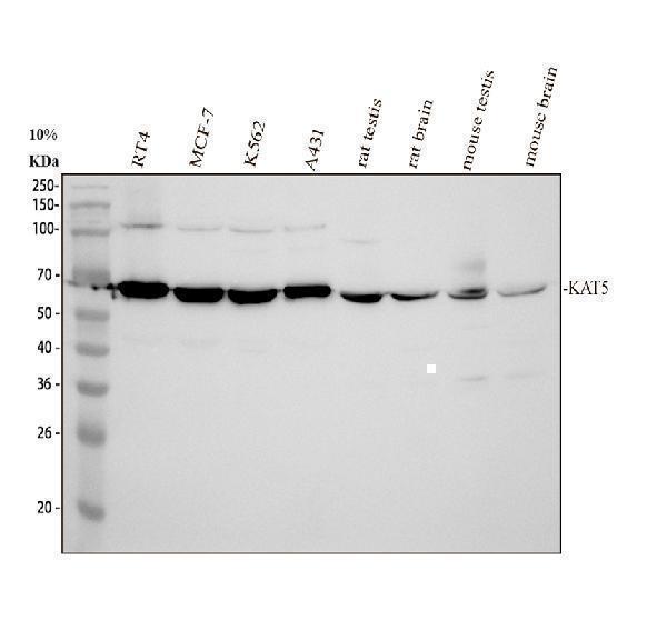
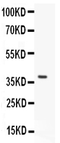
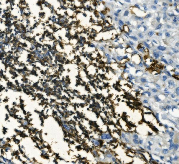
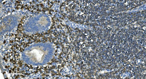
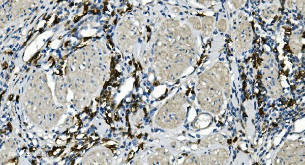
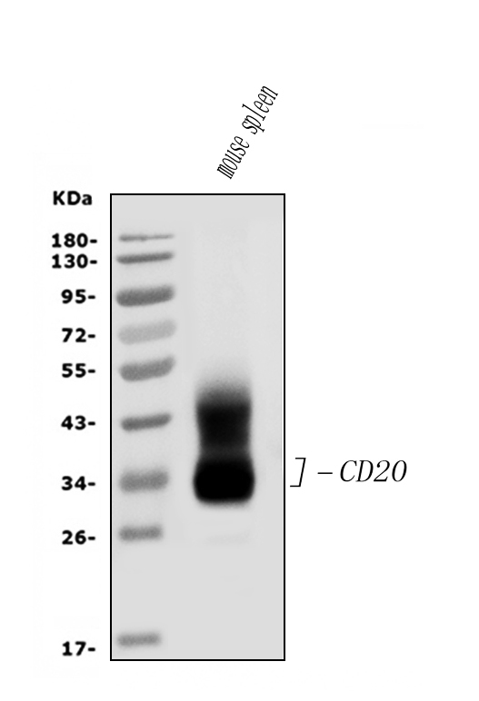
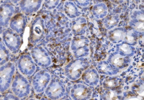
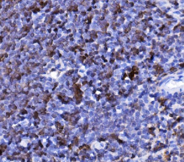
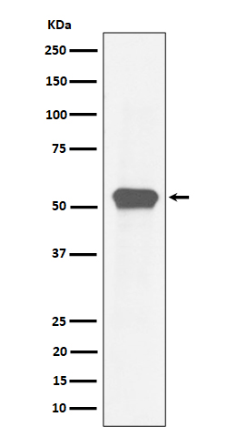
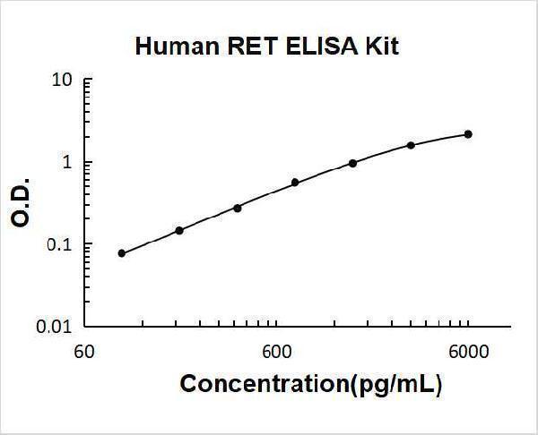
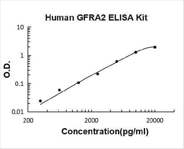
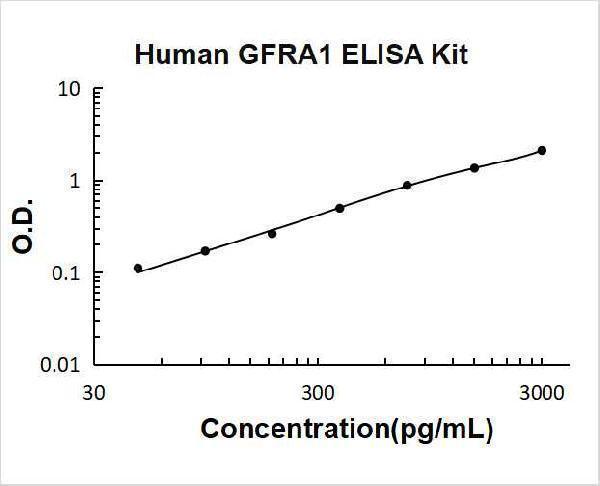
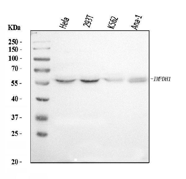
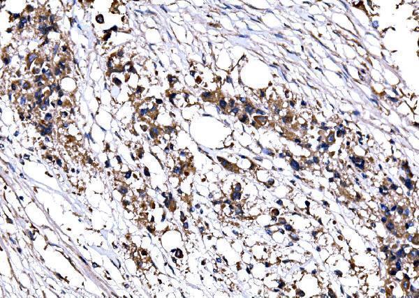
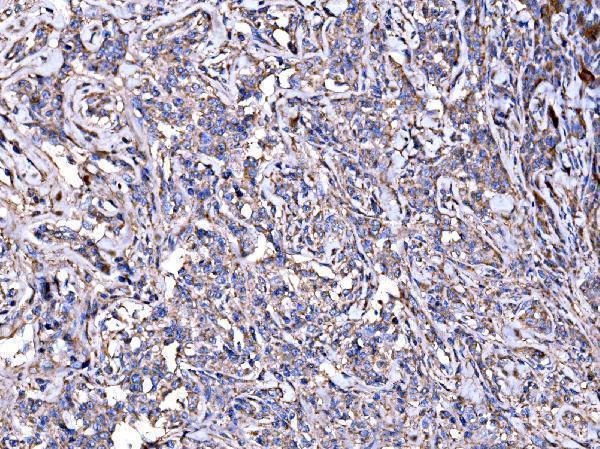
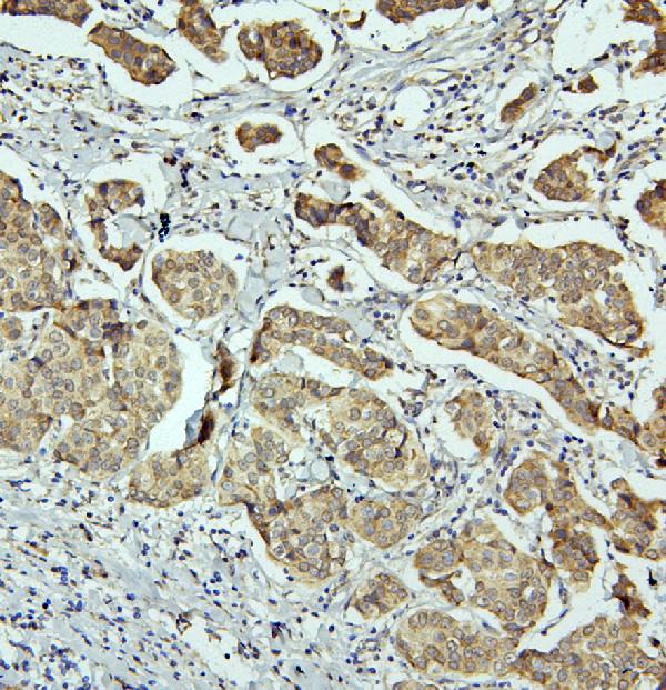
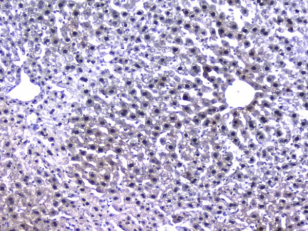
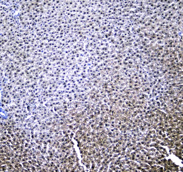
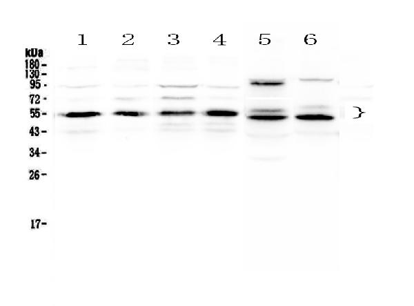
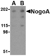
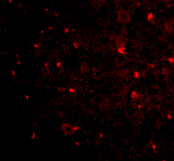
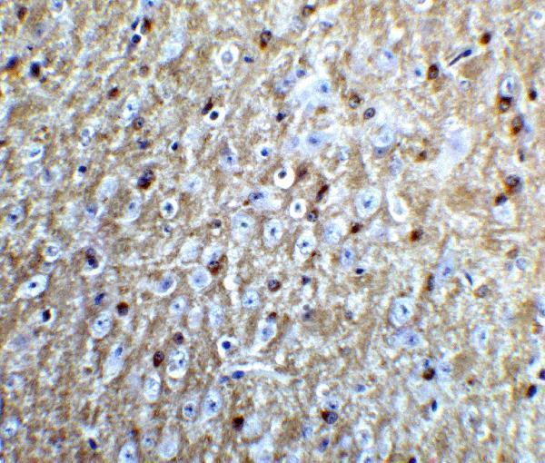
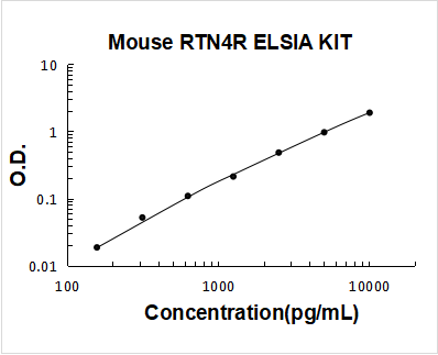
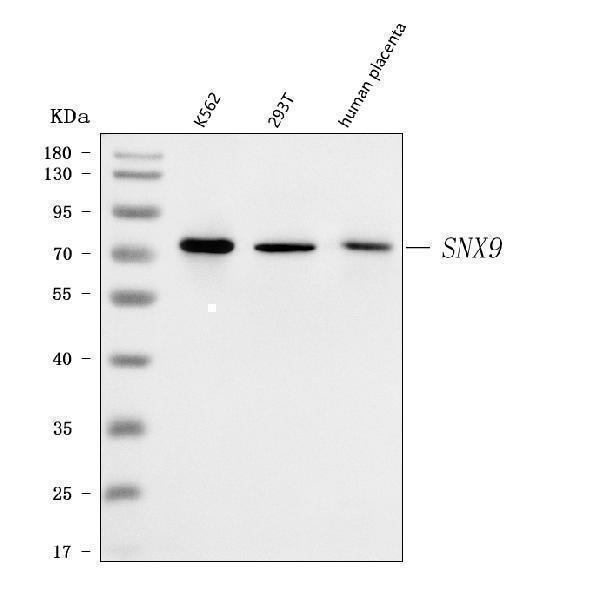
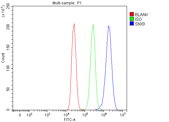
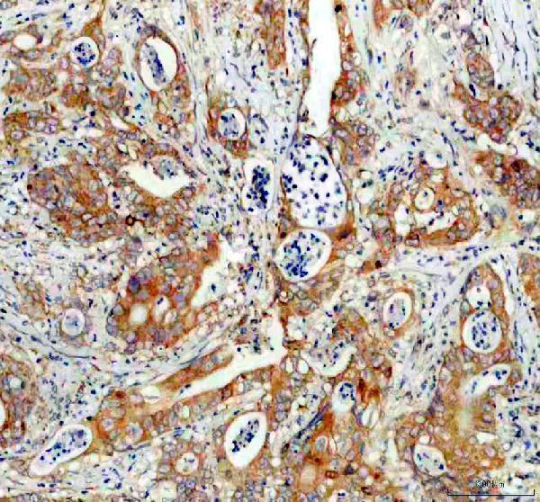
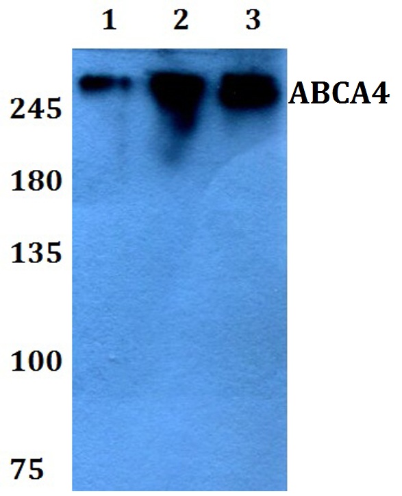
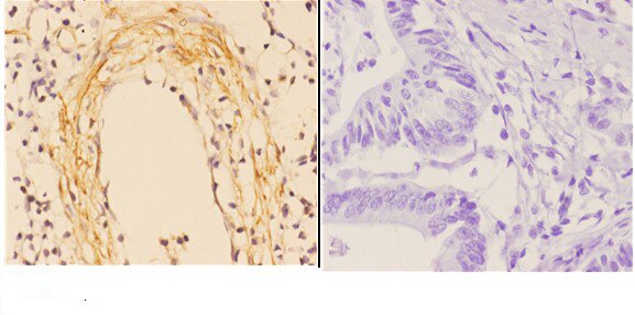
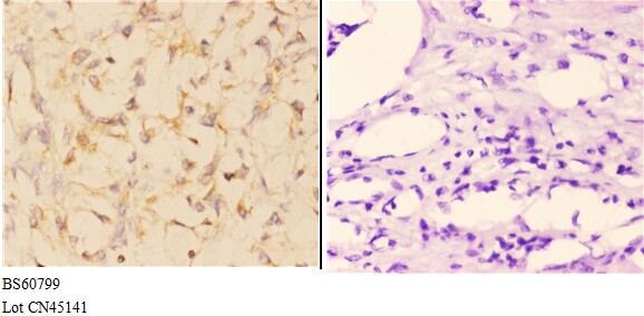
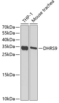
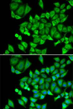
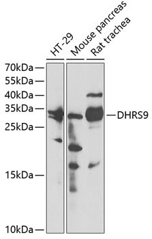
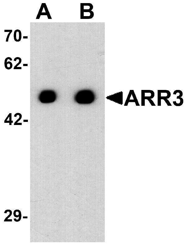
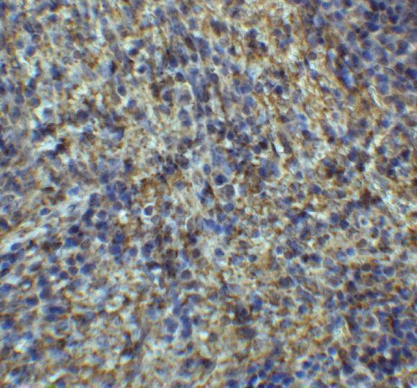
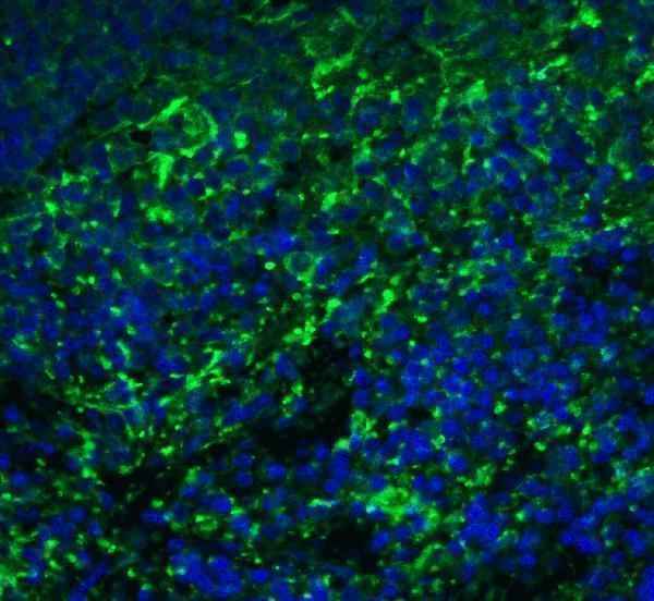
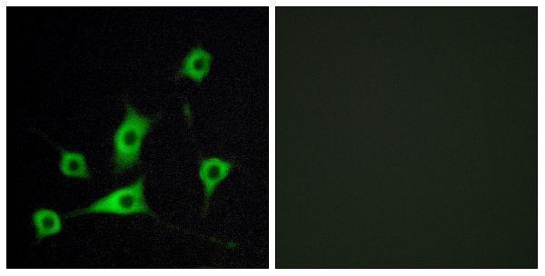
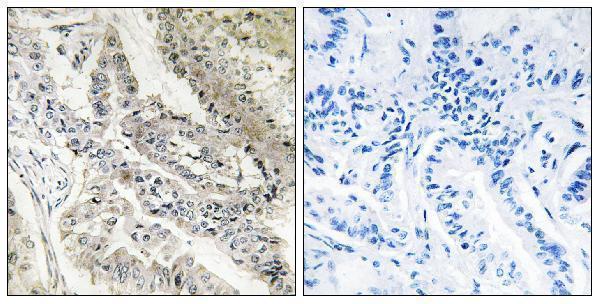
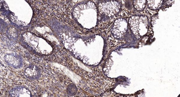
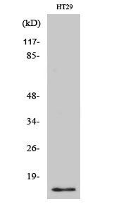
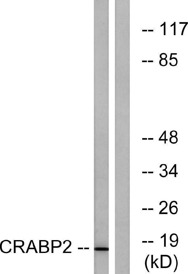
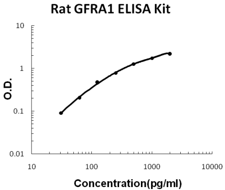
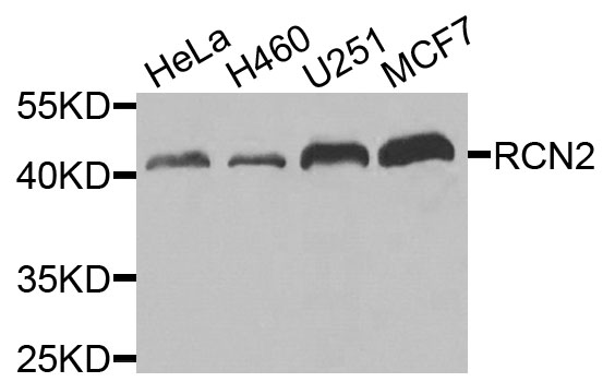
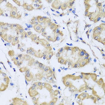
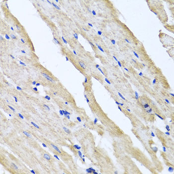
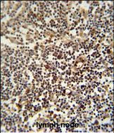
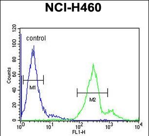
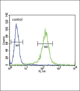
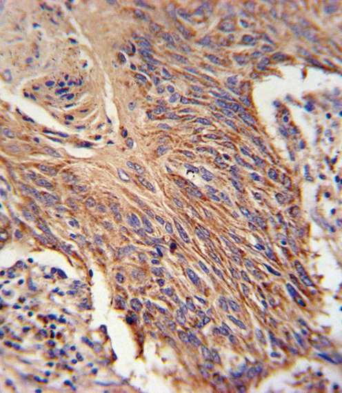
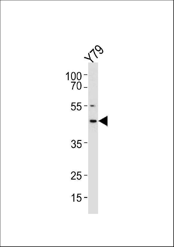
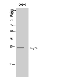
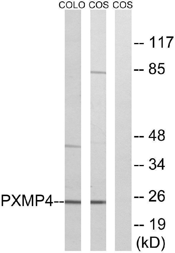
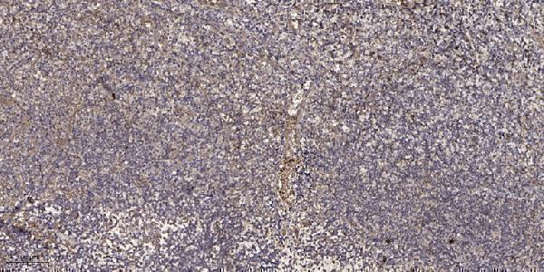
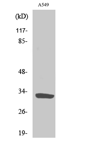
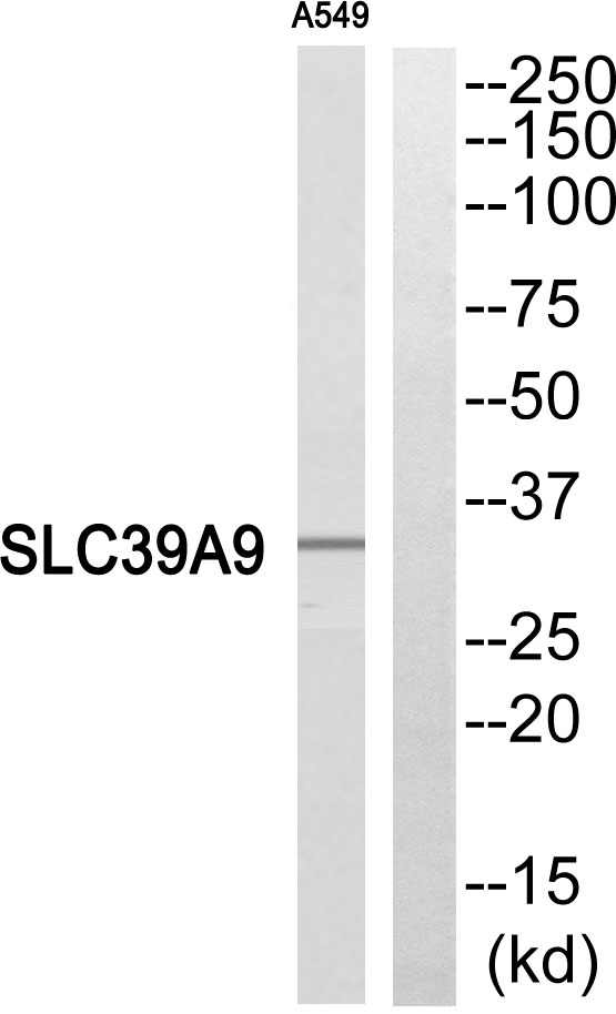
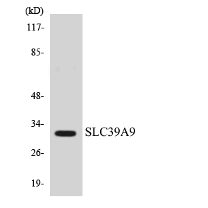
Many biological assays use antibodies to detect RET, such as Western Blot (WB), ELISA, Flow Cytometry, Immunocytochemistry (IHC). These antibodies can be polyclonal or monoclonal, and react with RET in Human, Mouse, Rat, and many other animal samples. Boster Bio use mainly mouse and rabbit to develop RET antibodies...
We validate the specificity of these antibodies to RET by testing them on tissues known to express RET positively and negatively. Browse below to find the RET antibody that suites your experiment. We have 100 of these antibodies and many publications and validation images.
If you cannot find antibodies that fit your needs, contact us for making custom antibodies. We have a full suite of custom antibody services covering from research to diagnostic and therapeutic applications.
Facts about Proto-oncogene tyrosine-protein kinase receptor Ret.
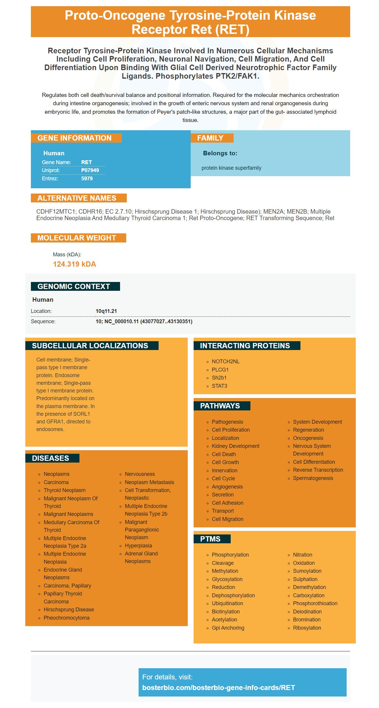
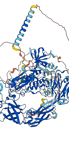
Proto-oncogene tyrosine-protein kinase receptor Ret (RET)
Receptor tyrosine-protein kinase involved in numerous cellular mechanisms including cell proliferation, neuronal navigation, cell migration, and cell differentiation upon binding with glial cell derived neurotrophic factor family ligands. Phosphorylates PTK2/FAK1.
Regulates both cell death/survival balance and positional information. Required for the molecular mechanics orchestration during intestine organogenesis; involved in the growth of enteric nervous system and renal organogenesis during embryonic life, and promotes the formation of Peyer's patch-like structures, a major part of the gut- associated lymphoid tissue.
Gene Information
Human
Gene Name:
RET
Uniprot:
P07949
Entrez:
5979
Family
Belongs to:
protein kinase superfamily
Alternative Names
CDHF12MTC1; CDHR16; EC 2.7.10; Hirschsprung disease 1; Hirschsprung disease); MEN2A; MEN2B; multiple endocrine neoplasia and medullary thyroid carcinoma 1; ret proto-oncogene; RET transforming sequence; Ret
Molecular Weight
Mass (kDA): 124.319 kDA
Genomic Context
Human
Location:
10q11.21
Sequence:
10; NC_000010.11 (43077027..43130351)
Subcellular Localizations
Cell membrane; Single-pass type I membrane protein. Endosome membrane; Single-pass type I membrane protein. Predominantly located on the plasma membrane. In the presence of SORL1 and GFRA1, directed to endosomes.
Diseases
Neoplasms
Carcinoma
Thyroid Neoplasm
Malignant Neoplasm Of Thyroid
Malignant Neoplasms
Medullary Carcinoma Of Thyroid
Multiple Endocrine Neoplasia Type 2a
Multiple Endocrine Neoplasia
Endocrine Gland Neoplasms
Carcinoma, Papillary
Papillary Thyroid Carcinoma
Hirschsprung Disease
Pheochromocytoma
Nervousness
Neoplasm Metastasis
Cell Transformation, Neoplastic
Multiple Endocrine Neoplasia Type 2b
Malignant Paraganglionic Neoplasm
Hyperplasia
Adrenal Gland Neoplasms
Interacting Proteins
NOTCH2NL
PLCG1
Sh2b1
STAT3
Pathways
Pathogenesis
Cell Proliferation
Localization
Kidney Development
Cell Death
Cell Growth
Innervation
Cell Cycle
Angiogenesis
Secretion
Cell Adhesion
Transport
Cell Migration
System Development
Regeneration
Oncogenesis
Nervous System Development
Cell Differentiation
Reverse Transcription
Spermatogenesis
PTMs
Phosphorylation
Cleavage
Methylation
Glycosylation
Reduction
Dephosphorylation
Ubiquitination
Biotinylation
Acetylation
Gpi Anchoring
Nitration
Oxidation
Sumoylation
Sulphation
Demethylation
Carboxylation
Phosphorothioation
Deiodination
Bromination
Ribosylation
For details, visit: bosterbio.com/bosterbio-gene-info-cards/RET
References
PMID: 2660074 by Takahashi M.; Isolation of ret proto-oncogene cDNA with an amino-terminal signal sequence.
PMID: 3078962 by Takahashi M., et al. Cloning and expression of the ret proto-oncogene encoding a tyrosine kinase with two potential transmembrane domains.
49 Publications of Boster RET Antibodies & ELISA Kits
PubMed ID: PMID:27114982, Effect of Omega-3 Supplementation on Lipocalin 2 and Retinol-Binding Protein 4 i...
PubMed ID: PMID:24995117, Ulinastatin attenuates cerebral ischemia-reperfusion injury in rats