This website uses cookies to ensure you get the best experience on our website.
- Table of Contents
and ELISA kits, proteins related to neuroscience research.
Neuroscience research investigates how the nervous system develops, functions, adapts, and degenerates across the brain, spinal cord, peripheral nerves, and neural model systems. Antibodies are essential tools in this field because they help researchers detect neuronal identity markers, glial and neuroimmune responses, synaptic proteins, signaling pathway nodes, and disease-associated biomarkers in both tissue and cell-based assays. In spatial biology workflows, IHC and IF are widely used to localize neurons, astrocytes, microglia, oligodendrocytes, and pathology-associated proteins in tissue sections. In mechanistic studies, Western blot supports pathway validation, synaptic protein analysis, and neurodegeneration-related readouts, while ELISA helps quantify cytokines, neurotrophic factors, and injury-associated soluble markers. Flow cytometry can be used to profile immune and neural cell populations in neuroinflammation and translational neuroscience studies. This neuroscience antibodies hub helps researchers find neuroscience-related biomarkers, disease-area antibody resources, cell-type entry points, pathway maps, and method guides for IHC, Western blot, ELISA, and flow cytometry workflows.
Neuroscience biomarker workflows often focus on neuronal identity, synaptic organization, glial activation, neuroinflammation, axonal injury, and neurodegeneration-associated protein changes. The targets below are commonly used across neurobiology, neurodegeneration, neuroinflammation, and translational brain research.
PA1239
MA1045
M00139
| Protein Name | Gene Name | Function |
|---|---|---|
| Brain-Derived Neurotrophic Factor (BDNF) | BDNF | Supports neuronal survival, growth, synaptic plasticity, and circuit adaptation. |
| Amyloid-beta / Amyloid Precursor Protein | APP | Central to amyloid processing and widely studied in Alzheimer’s disease and neurodegeneration. |
| Tau Protein | MAPT | Microtubule-associated protein linked to axonal stability, tau pathology, and neurodegenerative disease. |
| Glial Fibrillary Acidic Protein (GFAP) | GFAP | Canonical astrocyte marker used to assess gliosis, astrocyte activation, and neural injury response. |
| Apolipoprotein E (APOE) | APOE | Lipid transport and neurodegeneration-associated marker strongly linked to Alzheimer’s disease risk. |
| Neuron-Specific Enolase (NSE) | ENO2 | Neuronal marker frequently used in neural injury and neuroendocrine-related studies. |
| Neurofilament Light Chain (NfL) | NEFL | Axonal structural marker used in neurodegeneration and neuronal damage assessment. |
| S100B | S100B | Glial and injury-associated calcium-binding protein used in brain damage and neuroinflammation research. |
| Synaptophysin | SYP | Synaptic vesicle marker used to assess synapse density and neuronal connectivity. |
| Ubiquitin C-Terminal Hydrolase L1 (UCHL1) | UCHL1 | Neuron-enriched protein involved in protein turnover and neuronal maintenance. |
| Synaptosomal-Associated Protein 25 (SNAP-25) | SNAP25 | Key synaptic protein that supports neurotransmitter release and synaptic function. |
| Triggering Receptor Expressed on Myeloid Cells 2 (TREM2) | TREM2 | Microglia-associated receptor involved in injury sensing, phagocytosis, and neuroimmune regulation. |
| NOD-Like Receptor Protein 3 (NLRP3) | NLRP3 | Inflammasome component linked to neuroinflammation and injury-associated immune activation. |
| Postsynaptic Density Protein 95 (PSD-95) | DLG4 | Postsynaptic scaffold protein used in synapse organization and plasticity studies. |
| Matrix Metallopeptidase 9 (MMP-9) | MMP9 | Extracellular matrix remodeling factor associated with blood-brain barrier integrity and neuroinflammatory change. |
| Interleukin-6 (IL-6) | IL6 | Proinflammatory cytokine frequently measured in neuroinflammation and neural injury studies. |
| Interleukin-1 Beta (IL-1β) | IL1B | Inflammatory cytokine linked to neuroimmune activation, CNS injury, and disease progression. |
| Oxytocin | OXT | Neuropeptide involved in neural signaling, social behavior, and neurodevelopment-related studies. |
Use IHC to visualize neurons, astrocytes, microglia, oligodendrocytes, and disease-associated proteins in brain and spinal cord tissue. This is a core method for spatial neuroscience, neuropathology, and neurodegeneration studies.
Explore IHC resource centerWestern blot is commonly used to confirm expression of synaptic proteins, signaling nodes, axonal markers, and neurodegeneration-associated targets. It is especially useful for pathway validation and disease model comparison.
Explore Western blot resource centerUse ELISA to quantify inflammatory cytokines, neurotrophic proteins, injury-associated molecules, and soluble markers in neural tissue lysates, serum, CSF, or in vitro neuroscience models.
Explore ELISA resource centerFlow cytometry supports profiling of immune-related cell states, microglia-associated populations, and marker-defined cell subsets in neuroinflammation, injury, and translational neuroscience workflows.
Explore flow cytometry resource centerMany neuroscience workflows start with major CNS disease contexts such as Alzheimer’s disease, Parkinson’s disease, multiple sclerosis, and epilepsy. These studies frequently track neuronal injury, synaptic damage, glial activation, tau or amyloid-related pathology, and tissue-level disease progression using biomarker panels across IHC, WB, ELISA, and translational models.
Neuroscience research also extends into psychiatric and neurodevelopmental disease areas where neural signaling, synaptic regulation, immune crosstalk, and circuit dysfunction are major themes. These models are useful for connecting behavioral or developmental readouts with biomarker-level evidence from neuronal, glial, and inflammatory pathways.
A large part of neuroscience research focuses on how nervous tissue responds to damage and whether repair or regeneration is possible. These studies often involve axonal injury markers, glial transitions, blood-brain barrier disruption, demyelination-remyelination context, and developmental or stem-like programs that support recovery-oriented neuroscience models.
Many neuroscience studies begin with defining neuronal identity and synaptic organization. These workflows use neuronal and synapse-associated biomarkers to interpret brain-region specificity, connectivity, axonal integrity, and signaling changes across tissue, cell culture, and disease models.
Glial and neuroimmune context is essential for interpreting neuroinflammation, injury response, synaptic remodeling, and disease progression. These entry points are useful for biomarker panels involving astrocyte activation, microglial function, macrophage-like immune activity, and immune-cell-related CNS signaling.
Endothelial and barrier-associated biology is increasingly important in neuroscience, especially in neuroinflammation, neurovascular dysfunction, and injury models. These contexts help researchers interpret BBB integrity, leukocyte entry, tissue remodeling, and repair-associated environmental shifts in the nervous system.
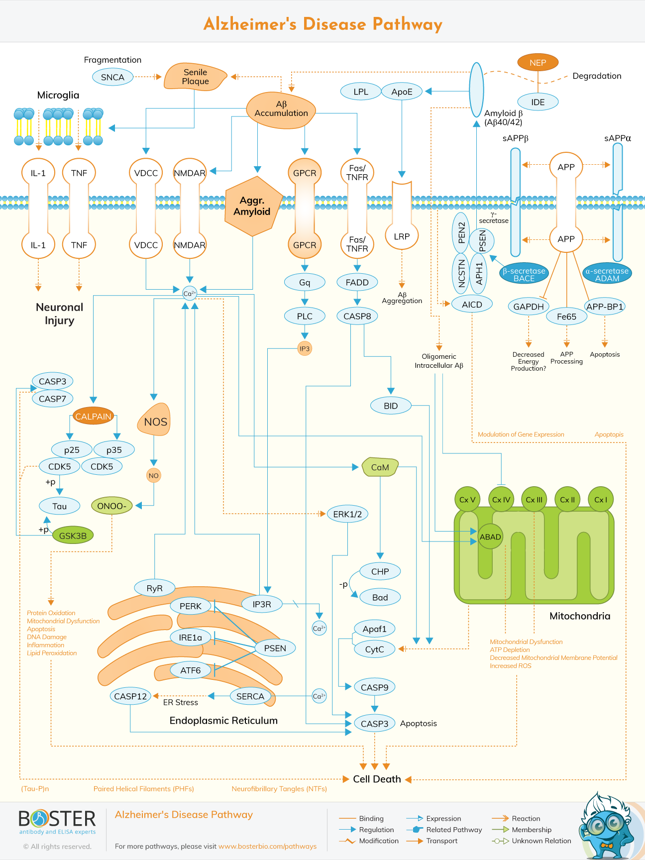
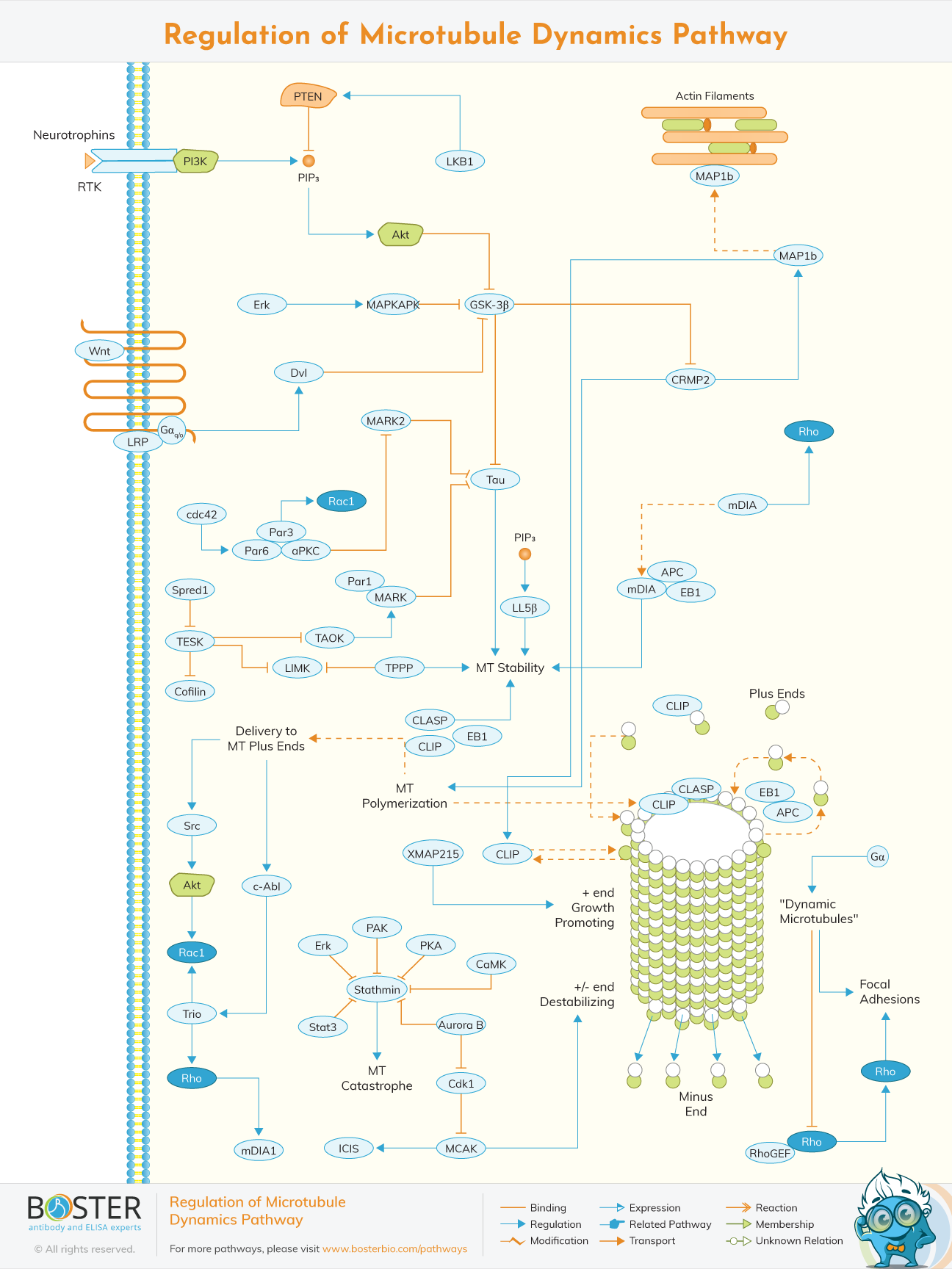
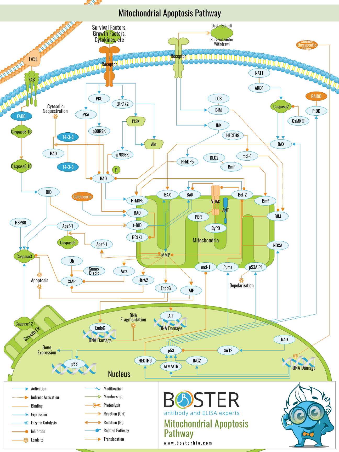
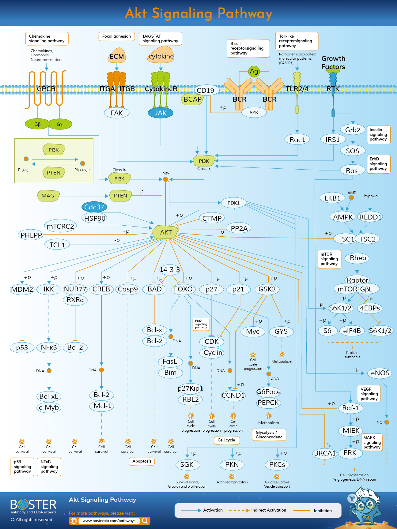
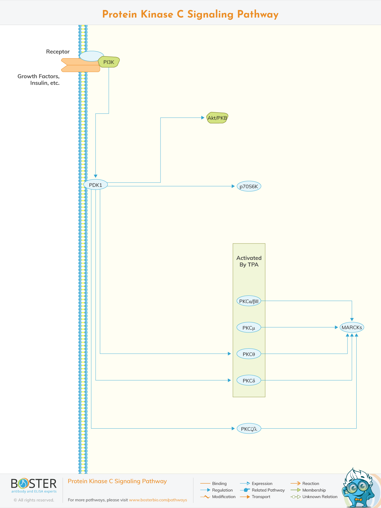
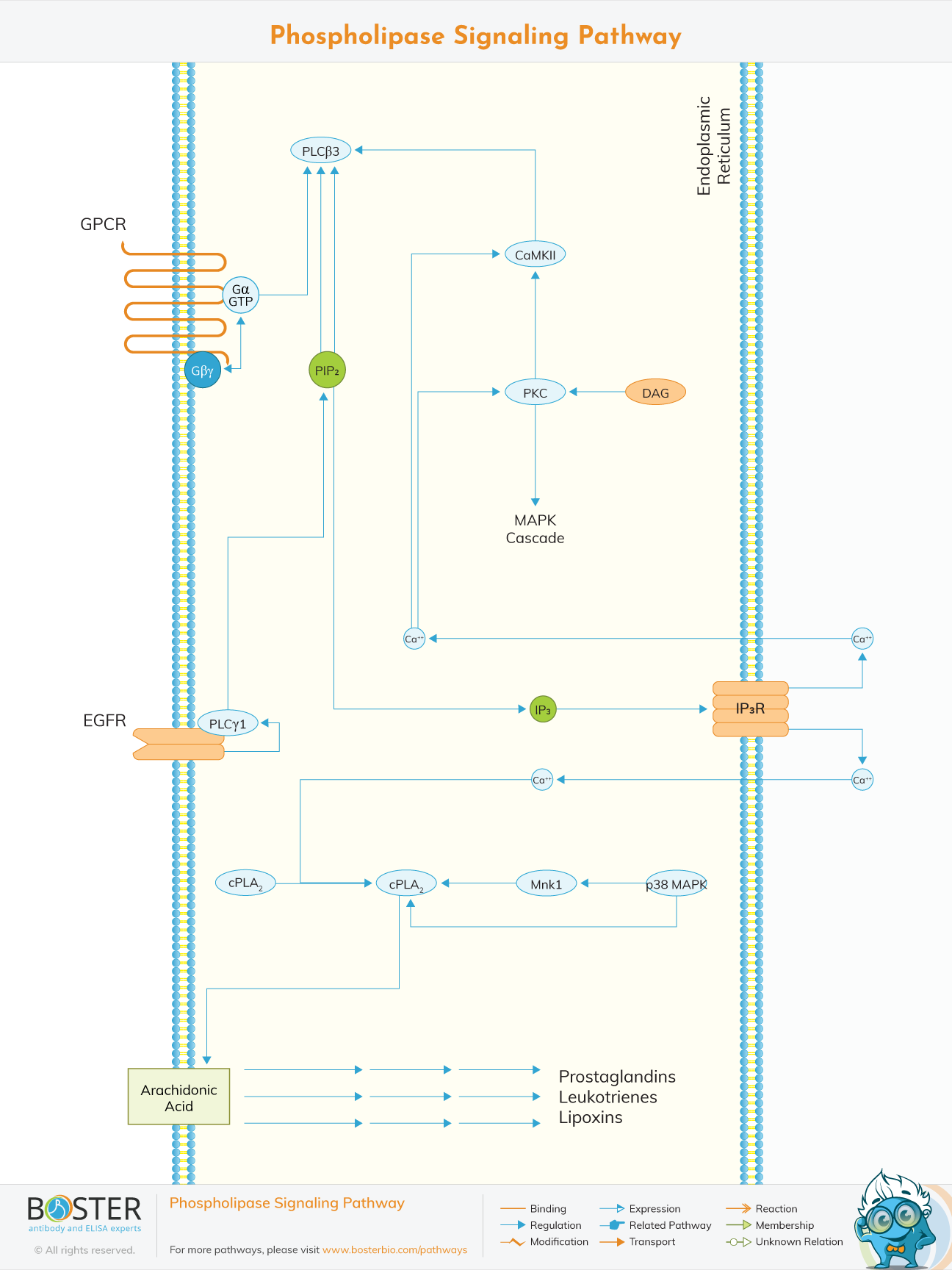
Pathway resources commonly used to interpret protein aggregation, disease progression, signaling dysregulation, and neural injury context.
Signaling maps relevant to neurotransmission, activity-dependent response, second messengers, and downstream neuronal pathway activation.
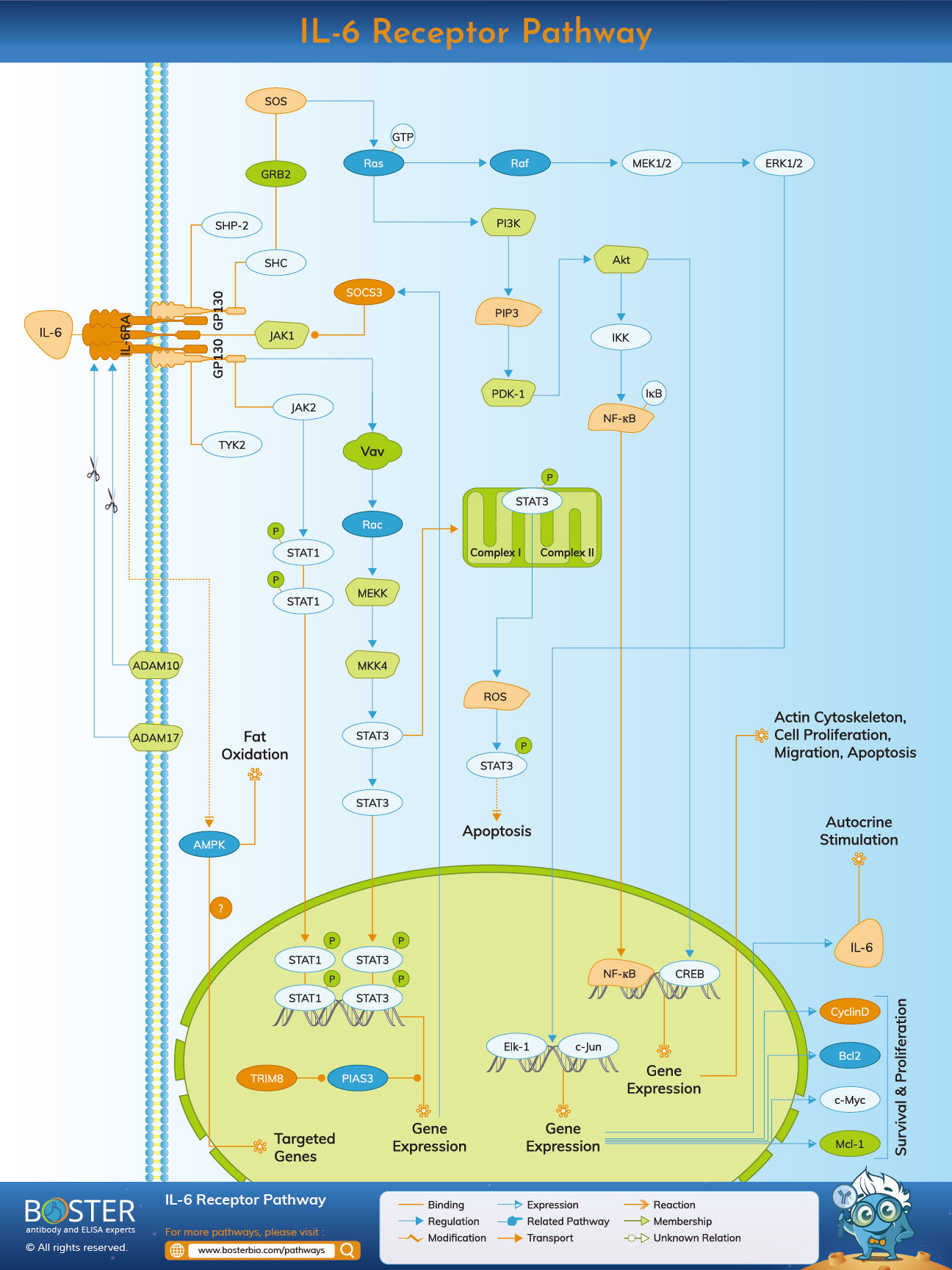
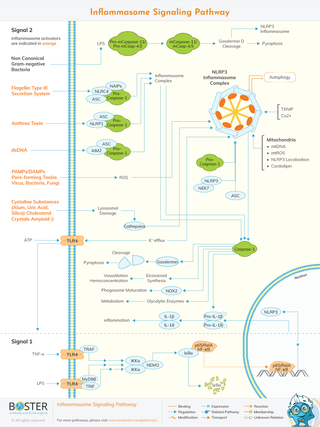
Pathway resources relevant to cytokine signaling, inflammasome activation, and immune-mediated neural damage or repair.
Synaptic function and plasticity are central to neuroscience because they connect molecular signaling to learning, memory, circuit adaptation, and behavioral output. Researchers in this area frequently study synaptic vesicle proteins, postsynaptic scaffolds, neurotransmitter release machinery, and activity-dependent signaling pathways to understand how neural networks strengthen, weaken, or reorganize over time. Biomarkers such as Synaptophysin, SNAP-25, PSD-95, and BDNF are commonly used across this space, together with pathway-level readouts linked to MAPK/ERK, Akt, calcium signaling, and cytoskeletal remodeling.
Neuroinflammation has become a major focus in neuroscience because immune signaling is deeply involved in neural injury, neurodegeneration, autoimmunity, and repair. Studies in this area often track astrocyte reactivity, microglial activation, inflammasome signaling, cytokine release, endothelial dysfunction, and blood-brain barrier disruption. Biomarkers such as GFAP, TREM2, NLRP3, IL-6, IL-1β, S100B, and MMP9 are frequently used to interpret glial state changes and inflammatory progression in CNS disease models.
Neurodegeneration research focuses on how neurons lose function, accumulate damage, and eventually die in disorders such as Alzheimer’s disease, Parkinson’s disease, and related CNS conditions. A major theme is the study of protein misfolding, aggregation, axonal injury, synaptic loss, and failure of cellular maintenance pathways. APP, Tau, APOE, NEFL, UCHL1, and related biomarkers are commonly used to connect pathological protein changes with tissue damage, disease progression, and pathway dysregulation in translational neuroscience workflows.