This website uses cookies to ensure you get the best experience on our website.


- Table of Contents
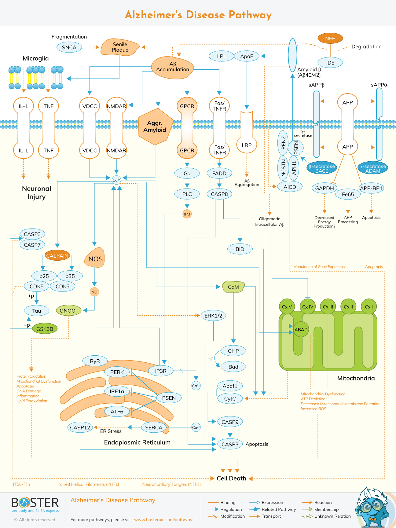
Interactive enrichment-driven map — APP processing, Aβ aggregation, tau hyperphosphorylation
Build your antibody bundle ↓| Target | SKU | Reactivity | Application | Size | CheckPlease select at least 4 targets. |
|---|---|---|---|---|---|
| SNCA | A00215-3 | Human, Mouse, Rat | ELISA, Flow Cytometry, IF, IHC, ICC, WB | 100 μg/vial | |
| APP (Senile Plaque) | A00081-3 | Human | IHC, WB | 100 μg/vial | |
| IL1B (IL-1) | A00101 | Human, Mouse, Rat | WB | 100 μg/vial | |
| TNF | A00002-2 | Mouse | WB | 100 μg/vial | |
| CACNA1C (VDCC) | No PicoBand® antibody for this target — null | ||||
| GRIN2B (NMDAR) | PB9373 | Mouse, Rat | WB | 100 μg/vial | |
| GRM1 (GPCR) | A03049-4 | Human, Mouse, Rat | WB, IHC, Flow Cytometry, ELISA | 100 μg/vial | |
| FAS (Fas/TNFR) | A00055 | Mouse, Rat | ELISA, Flow Cytometry, WB | 100 μg/vial | |
| LRP1 (LRP) | No PicoBand® antibody for this target — null | ||||
| LPL | No PicoBand® antibody for this target — null | ||||
| APOE (ApoE) | A00015-5 | Human | ELISA, Flow Cytometry, IF, IHC, ICC, WB | 100 µg/vial | |
| MME (NEP) | PB9058 | Human, Rat | Flow Cytometry, IHC, WB | 100 μg/vial | |
| IDE | A01358-2 | Human, Mouse, Rat | ELISA, Flow Cytometry, IHC, ICC, WB | 100 μg/vial | |
| ADAM10 (α-secretase ADAM) | A00566-1 | Human, Mouse, Rat | WB, ELISA | 100 μg/vial | |
| BACE1 (β-secretase BACE) | A00322-3 | Human, Mouse, Rat | WB, ELISA | 100 μg/vial | |
| GAPDH | A00227-1 | Chicken, Human, Monkey, Mouse, Rat, Zebrafish | IF, IHC, ICC, WB, Flow Cytometry | 100 μg/vial | |
| APBB1 (Fe65) | RP1059 | Human, Mouse | WB | 100 μg/vial | |
| NAE1 (APP-BP1) | PB9339 | Human, Mouse, Rat | WB | 100 μg/vial | |
| PSENEN (PEN2) | No PicoBand® antibody for this target — null | ||||
| PSEN1 (PSEN) | A00138-3 | Human, Mouse, Rat | ELISA, Flow Cytometry, IF, ICC, WB | 100 μg/vial | |
| NCSTN | A03144-1 | Human, Mouse, Rat | WB, ICC, IF, ELISA | 100 μg/vial | |
| APH1A (APH1) | A04859-1 | Human, Mouse, Rat | Flow Cytometry, IHC, WB | 100 μg/vial | |
| CASP3 (CASP3 (apop)) | A00334-2 | Human | WB, ICC, IF, Flow Cytometry | 100 μg/vial | |
| CASP7 | A01044-2 | Human, Rat | ELISA, Flow Cytometry, IF, IHC, ICC, WB | 100 μg/vial | |
| CAPN1 (CALPAIN) | PA1364 | Human, Mouse, Rat | IF, IHC, IHC-F, ICC, WB | 100 μg/vial | |
| CDK5R1 (p25) | No PicoBand® antibody for this target — null | ||||
| CDK5 | A00511-1 | Human, Mouse, Rat | ELISA, IHC, WB | 100 μg/vial | |
| MAPT (Tau) | A00097-3 | Human, Mouse, Rat | ELISA, IF, IHC, ICC, WB | 100 μg/vial | |
| GSK3B | A00791-3 | Human, Mouse, Rat | ELISA, IF, IHC, ICC, WB | 100 μg/vial | |
| NOS1 (NOS) | A01070 | Human, Mouse, Rat | WB | 100 μg/vial | |
| NOS2 (ONOO-) | A00368 | Human | IHC, WB | 100 μg/vial | |
| GNAQ (Gq) | PB9707 | Human, Mouse, Rat | Flow Cytometry, IF, IHC, ICC, WB | 100 μg/vial | |
| PLCB1 (PLC) | A02817-1 | Human, Mouse, Rat | WB, ELISA | 100 µg/vial | |
| ITPR1 (IP3) | PB9225 | Human, Mouse, Rat | Flow Cytometry, IF, IHC, ICC, WB | 100 μg/vial | |
| FADD | PA1464 | Human, Mouse, Rat | WB | 100 μg/vial | |
| CASP8 | A00042 | Human, Mouse, Rat | Flow Cytometry, IHC, IHC-F, ICC, WB | 100 μg/vial | |
| BID | A00730 | Mouse, Rat | Flow Cytometry, IHC, WB | 100 μg/vial | |
| CALM1 (CaM) | No PicoBand® antibody for this target — null | ||||
| MAPK1 (ERK1/2) | PA1310 | Human, Mouse, Rat | WB | 100 μg/vial | |
| CHP1 (CHP) | A04737-3 | Human, Mouse, Rat | WB, ICC, IF, Flow Cytometry | 100 μg/vial | |
| BAD (Bad) | A03520-3 | Human, Mouse | WB, Flow Cytometry | 100 μg/vial | |
| APAF1 (Apaf1) | A00889-2 | Human | WB, Flow Cytometry, ELISA | 100 μg/vial | |
| CYCS (CytC) | A03529 | Human, Mouse, Rat | IHC, WB | 100 μg/vial | |
| CASP9 | A00080-5 | Human | WB, IHC, ICC, IF, Flow Cytometry, ELISA | 100 μg/vial | |
| RYR2 (RyR) | A00155-2 | Human | WB, IHC, ELISA | 100 μg/vial | |
| EIF2AK3 (PERK) | A01992-2 | Human | ELISA, IHC, WB | 100 μg/vial | |
| ERN1 (IRE1a) | A00683-1 | Human, Mouse, Rat | ELISA, Flow Cytometry, IF, ICC, WB | 100 μg/vial | |
| ATF6 | A00655 | Human, Mouse, Rat | IHC, WB | 100 μg/vial | |
| CASP4 (CASP12) | A02941-1 | Human | ELISA, Flow Cytometry, IF, IHC, ICC, WB | 100 μg/vial | |
| NDUFS1 (Cx I) | A04920-1 | Human, Mouse, Rat | ELISA, Flow Cytometry, IHC, WB | 100 μg/vial | |
| SDHA (Cx II) | A01753 | Human, Mouse, Rat | Flow Cytometry, IF, ICC, WB | 100 μg/vial | |
| UQCRC1 (Cx III) | No PicoBand® antibody for this target — null | ||||
| MT-CO1 (Cx IV) | PA1317 | Human | WB | 100 μg/vial | |
| NDUFA9 (Cx V) | A07420-3 | Human, Mouse, Rat | ELISA, Flow Cytometry, IF, IHC, ICC, WB | 100 µg/vial | |
| HSD17B10 (ABAD) | PB9336 | Human | Flow Cytometry, IF, IHC, ICC, WB | 100 μg/vial | |
| ATP2A2 (SERCA) | No PicoBand® antibody for this target — null | ||||
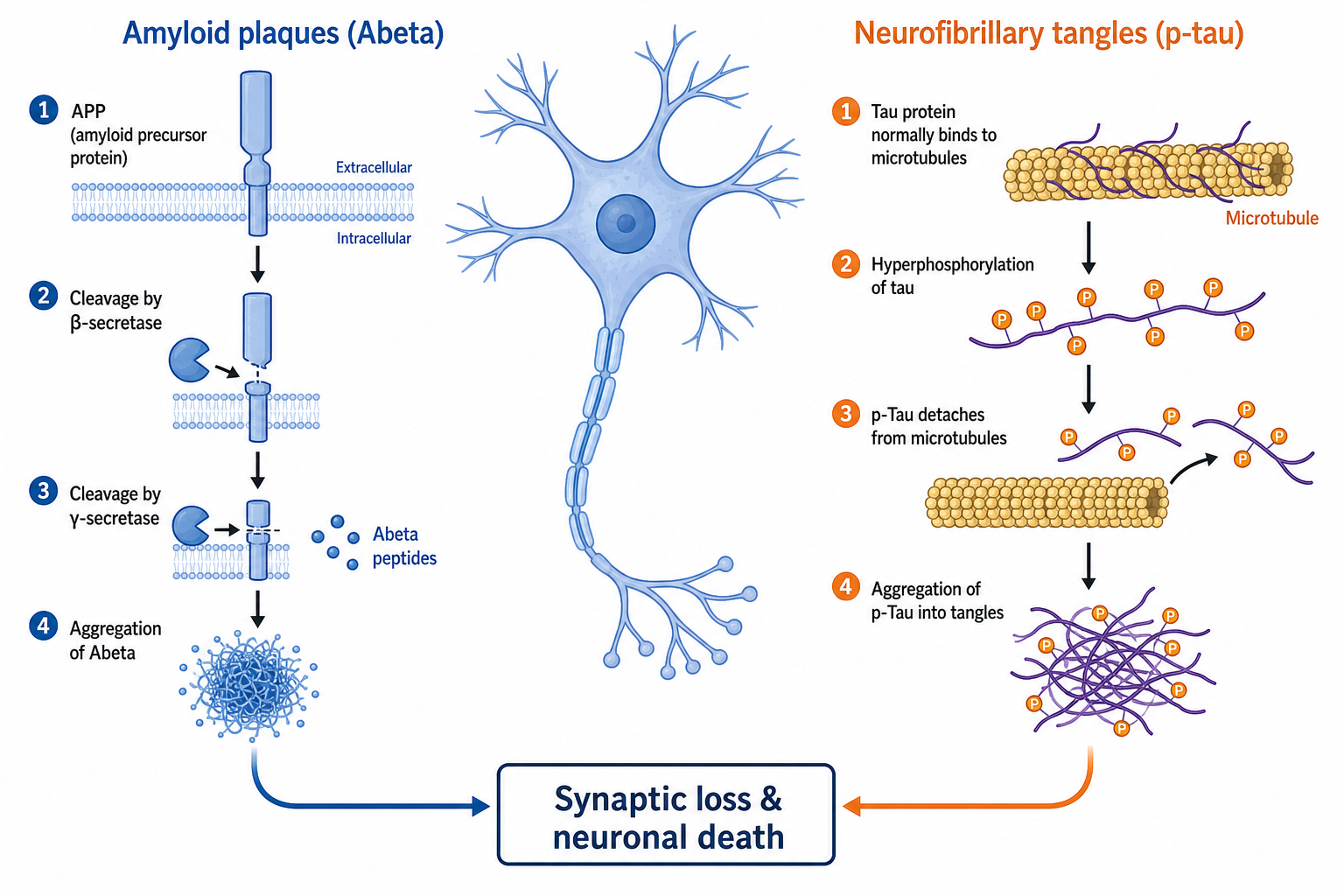
The Alzheimer's disease pathway is the network of molecular events that drives the irreversible decline in cognitive and behavioral ability that defines Alzheimer's disease (AD). It is built on two convergent protein-misfolding axes: the amyloid secretase pathway, which generates and aggregates β-amyloid (Aβ) peptides into extracellular neuritic plaques, and the tau pathway, in which hyperphosphorylated tau detaches from microtubules and assembles into intraneuronal neurofibrillary tangles (NFTs).
These two lesions do not act in isolation. Aβ oligomers trigger inflammatory cascades, dysregulated calcium handling, oxidative stress, and neuronal apoptosis, and they promote tau phosphorylation. Upstream kinases — most notably GSK3β — and the Wnt signaling axis sit above both lesions, linking impaired Aβ clearance, tau hyperphosphorylation, reduced neurogenesis, and amplified apoptosis into one self-reinforcing program.
Understanding the Alzheimer's disease signaling pathway is foundational for research in neurodegeneration, biomarker discovery, and the development of disease-modifying therapeutics that target the underlying biology rather than only the cholinergic symptoms.
What every researcher should know before designing an Alzheimer's-pathway experiment.
Soluble Aβ42 oligomers, not mature plaques, drive synaptotoxicity — triggering inflammation, calcium dysregulation, oxidative stress, and tau phosphorylation. The β-/γ-secretase processing balance is the molecular control point of the amyloid secretase pathway.
Tau has ~79 serine/threonine phosphorylation sites. The phosphorylation/dephosphorylation equilibrium — governed by GSK3β, CDK5, MAPK and phosphatases PP1/PP2A/PP5 — decides whether microtubules stay stable or collapse into neurofibrillary tangles.
Hyperactive GSK3β simultaneously hyperphosphorylates tau, promotes amyloidogenic APP cleavage, suppresses neurogenesis, and amplifies apoptosis. Wnt signaling normally restrains it — Wnt loss in AD removes that brake.
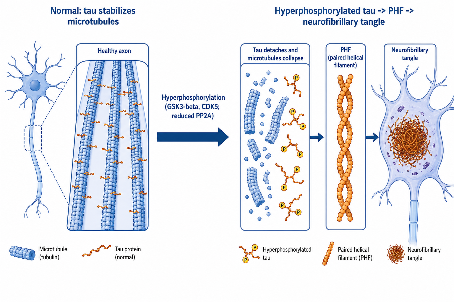
Tau is a microtubule-associated protein concentrated in the central and peripheral nervous system and a key part of the neuronal cytoskeleton. By interacting with α- and β-tubulin it stabilizes microtubules, which are essential for neuronal structure, synaptic plasticity, and axonal transport.
Abnormal hyperphosphorylation impairs tubulin binding, damages microtubule architecture, disrupts axonal transport, and ultimately causes cytoskeletal breakdown and neuronal death.
A network of kinases and phosphatases sets the tau phosphorylation state:
Loss of phosphatase activity combined with kinase hyperactivity produces the pathologically hyperphosphorylated tau that seeds tangle formation.
Neurofibrillary tangles are intraneuronal lesions composed mainly of paired helical filaments (PHFs) of hyperphosphorylated tau:
Intraneuronal hyperphosphorylated tau is detectable in mild dementia even without β-amyloid pathology, suggesting tau dysregulation can be an early initiating event.
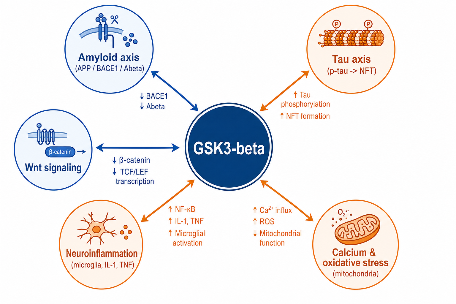
GSK3β is a hub linking the tau and amyloid axes:
Because GSK3β sits upstream of both lesions, it is one of the most studied convergence-node drug targets in the Alzheimer's disease pathway.
The Alzheimer's disease pathway is not a single linear chain. The amyloid and tau axes are coupled to each other and to the major stress- and death-signaling systems of the neuron. Mapping these connections prevents misinterpretation of experimental results and reveals where intervention is most tractable.
Aβ oligomers activate neuronal kinases — prominently GSK3β and CDK5 — that hyperphosphorylate tau. This is the molecular bridge between the two hallmark lesions: amyloid pathology accelerates tau pathology, while tau is required for Aβ-induced neurotoxicity. Targeting only one axis often leaves the other to drive progression.
Aβ deposits activate microglia and astrocytes, releasing IL-1β, TNF-α, and other mediators. This neuroinflammatory response further impairs Aβ clearance and amplifies neuronal injury, creating a feed-forward loop in which inflammation both responds to and aggravates the amyloid burden.
Aβ oligomers form membrane pores and dysregulate intracellular calcium, while mitochondrial dysfunction raises reactive oxygen species. Calcium dyshomeostasis and oxidative stress reinforce each other and feed back onto kinase activity, further driving tau phosphorylation and synaptic failure.
Wnt signaling normally inactivates GSK3β. Loss of Wnt tone in AD releases GSK3β, which simultaneously hyperphosphorylates tau, biases APP toward amyloidogenic cleavage, suppresses neurogenesis, and promotes apoptosis — making the Wnt/GSK3β node a single lever that moves both lesions at once.
The same amyloid and tau machinery produces different outcomes depending on which CNS cell type is involved. These are the most experimentally relevant contexts.
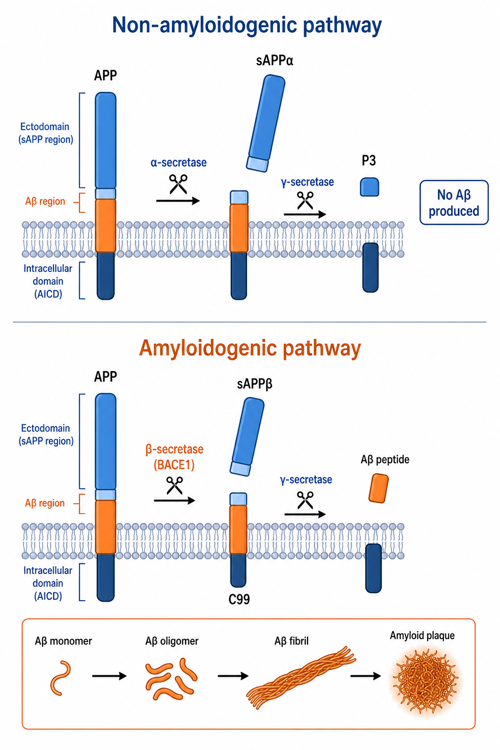
Amyloid precursor protein (APP) is an integral polytopic protein highly expressed in the central nervous system and present in peripheral epithelium and blood cells. APP is processed by two competing routes, and the balance between them determines whether toxic Aβ accumulates.
| Route | Initiating Enzyme | Fragments Produced | Outcome |
|---|---|---|---|
| Non-amyloidogenic | α-secretase (within the Aβ domain) | sAPPα (soluble N-terminal) + C83 | C83 cleaved by γ-secretase to the short P3 fragment; no intact Aβ — neuroprotective default route |
| Amyloidogenic | β-secretase (BACE1) | sAPPβ (shorter N-terminal) + C99 | C99 cleaved by γ-secretase to full-length Aβ (Aβ40 / Aβ42) |
| Aggregation | Concentration-driven (Aβ42 prone) | Monomers → dimers → oligomers → protofibrils → fibrils | Mature into diffuse, then neuritic, then senile amyloid plaques |
In the non-amyloidogenic pathway, α-secretase cleaves APP within the Aβ sequence, releasing sAPPα and the membrane-bound C83 fragment, which γ-secretase further trims to the innocuous P3 peptide. No intact Aβ is generated. Under physiological conditions this is the preferred route, and a balance is maintained between the small amount of Aβ produced and its clearance.
In the amyloidogenic pathway, β-secretase (BACE1) cleaves APP to sAPPβ and C99; γ-secretase then liberates the full-length Aβ peptide. Aβ40 and Aβ42 dominate in the brain, with Aβ42 the more aggregation-prone and neurotoxic species and the central driver of AD pathogenesis. Secreted Aβ monomers progressively assemble into dimers, oligomers, protofibrils, and fibrils that deposit and mature into amyloid plaques.
The soluble Aβ oligomers — not the mature plaques — are regarded as the most lethal species. They interact with neurons and glia to trigger inflammatory cascades, dysregulated calcium metabolism, oxidative stress, neuronal apoptosis, and tau phosphorylation. Clearance normally depends on Apolipoprotein E (ApoE) and the insulin-degrading enzyme (IDE); in disease, a metabolic shift toward amyloidogenic cleavage plus reduced clearance drives net Aβ accumulation.
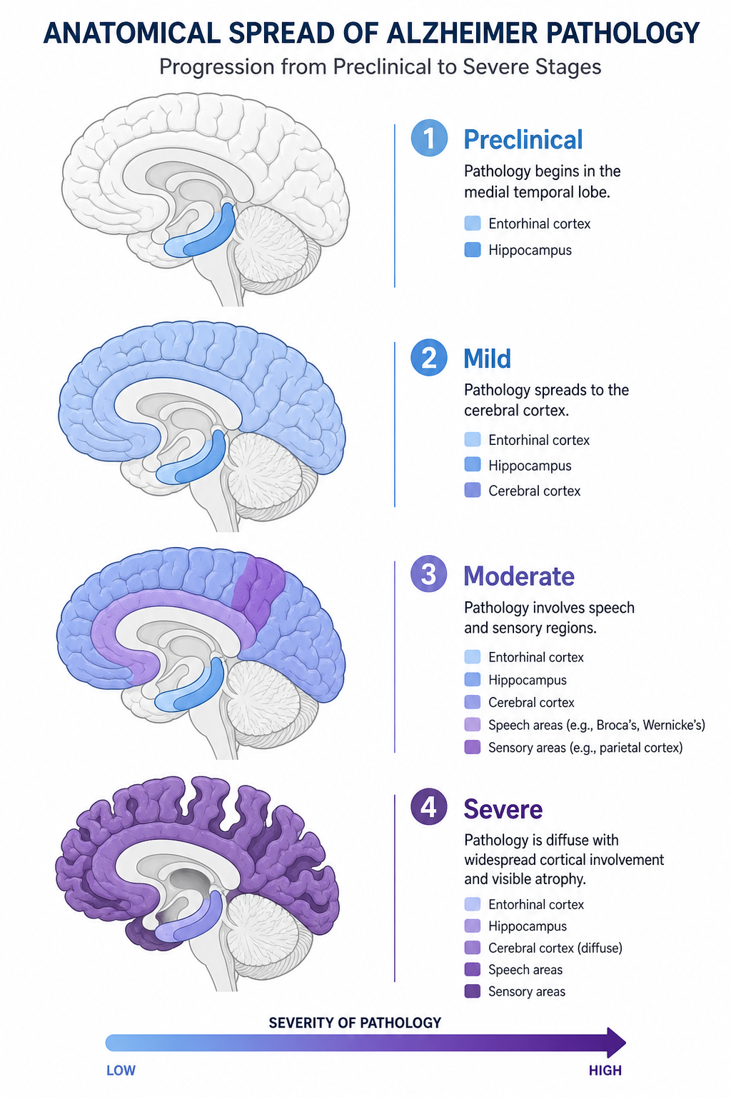
AD pathology begins years before symptoms and spreads through the brain in a stereotyped anatomical sequence, which is why pathway readouts are interpreted differently depending on disease stage.
The hallmark lesions — neuritic plaques rich in Aβ42 and neurofibrillary tangles of hyperphosphorylated tau, accompanied by gliosis and neuronal loss — accumulate in a sequence that mirrors symptom progression. Staging the dominant lesion in a model system is essential for choosing the right pathway readout.
Selecting the right readout for an Alzheimer's-pathway experiment requires knowing which gene belongs to which functional node. The table below maps key Alzheimer's disease pathway components to their function, standard detection method, and experimental context.
| Gene / Protein | Pathway Node | Function | Common Detection Method |
|---|---|---|---|
| APP | Amyloid substrate | Amyloid precursor protein; substrate for α-/β-/γ-secretase cleavage | Western blot, IHC, ELISA |
| BACE1 | β-secretase | Initiates amyloidogenic processing; rate-limiting for Aβ generation | Western blot, activity assay |
| PSEN1 (Presenilin-1) | γ-secretase | Catalytic core of γ-secretase; familial AD mutations shift Aβ42/Aβ40 ratio | Western blot, sequencing |
| Aβ42 (APP-derived peptide) | Toxic effector | Aggregation-prone amyloid peptide; primary plaque component | ELISA (CSF/plasma), IHC |
| MAPT (Tau) | Microtubule / NFT | Microtubule-associated protein; hyperphosphorylated form forms PHFs/NFTs | Western blot, IHC, ELISA |
| p-Tau (phospho-tau) | NFT marker | Hyperphosphorylated tau; correlates with tangle burden and progression | ELISA (CSF/plasma), IHC |
| GSK3B | Convergence kinase | Central tau kinase; promotes amyloidogenic APP cleavage and apoptosis | Western blot, activity assay |
| CDK5 | Tau kinase | Proline-directed kinase; tau hyperphosphorylation in AD cortex | Western blot, kinase assay |
| APOE | Aβ clearance / risk | ApoE4 isoform is the major late-onset AD risk allele; impairs Aβ clearance | Genotyping, ELISA, IHC |
| PP2A (PPP2CA) | Tau phosphatase | Major tau phosphatase; reduced activity in AD shifts tau to hyperphospho state | Western blot, phosphatase assay |
When designing an Alzheimer's-pathway experiment, CSF or plasma Aβ42 and phospho-tau are the most validated fluid biomarkers, while tau and p-tau IHC are the standard tissue readouts for tangle burden. For mechanistic work, GSK3β activity assays or BACE1 activity assays provide direct evidence of pathway engagement at the convergence and amyloidogenic-initiation steps respectively.
Common questions from researchers working with the Alzheimer's disease pathway.
The Alzheimer's disease pathway is the molecular network that produces the two hallmark lesions of AD: extracellular amyloid plaques from β-/γ-secretase processing of APP, and intraneuronal neurofibrillary tangles of hyperphosphorylated tau. Upstream kinases such as GSK3β and the Wnt axis link impaired Aβ clearance, tau hyperphosphorylation, neuroinflammation, and apoptosis into one self-reinforcing program.
For research it is relevant across neurodegeneration, biomarker discovery, and disease-modifying drug development. Knowing which axis (amyloid vs. tau) and which node is dominant in your model is essential for interpreting results correctly.
Both describe how APP is cleaved. In the non-amyloidogenic pathway, α-secretase cuts APP within the Aβ sequence to release sAPPα and C83; γ-secretase then trims C83 to the harmless P3 fragment — no intact Aβ is produced. This is the preferred physiological route.
In the amyloidogenic pathway, β-secretase (BACE1) cleaves APP to sAPPβ and C99; γ-secretase then liberates full-length Aβ (Aβ40/Aβ42). Aβ42 is the more aggregation-prone, neurotoxic species. A disease-associated shift toward amyloidogenic cleavage plus reduced ApoE/IDE-mediated clearance drives net Aβ accumulation.
The Alzheimer's disease KEGG map is catalogued under entry hsa05010 (Homo sapiens), freely accessible at the KEGG PATHWAY database (genome.jp/kegg/pathway.html). It covers APP processing by the secretases, Aβ aggregation, tau hyperphosphorylation, and the downstream calcium, mitochondrial, ER-stress, and apoptotic branches.
A complementary curated resource is "AlzPathway," a comprehensive Alzheimer's signaling map of roughly 1,347 molecules, 1,070 reactions, and 129 phenotypes across neurons, the blood-brain barrier, astrocytes, synapses, and microglia — useful for building targeted gene panels or interpreting pathway-enrichment results.
Alzheimer's disease drugs fall into two broad categories:
Botanical EGb 761 (Ginkgo biloba) is used in Europe for symptomatic relief — neuroprotective and antioxidant — but is not proven to halt progression to dementia.
The approach depends on which axis you are probing and the mechanistic depth required:
The sequence is still debated, and the evidence supports a coupled rather than strictly linear relationship. The amyloid cascade hypothesis places Aβ accumulation upstream, with Aβ oligomers triggering the kinase activity (GSK3β, CDK5) that hyperphosphorylates tau.
However, intraneuronal hyperphosphorylated tau is detectable in mild dementia even without β-amyloid pathology, and NFT spread correlates more tightly with cognitive decline than plaque load. This suggests tau dysregulation may be an early initiating event in some contexts, with Aβ over-secretion, oxidative stress, and inflammation acting as amplifiers. The practical implication: stage your model and measure both axes rather than assuming a fixed order.