This website uses cookies to ensure you get the best experience on our website.


- Table of Contents
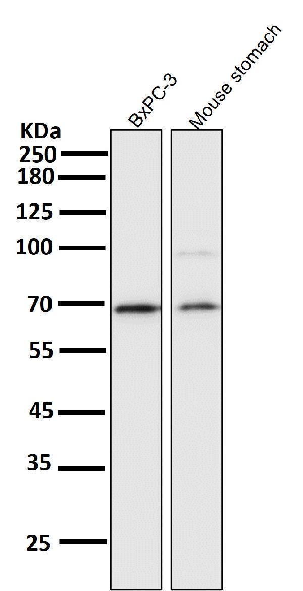
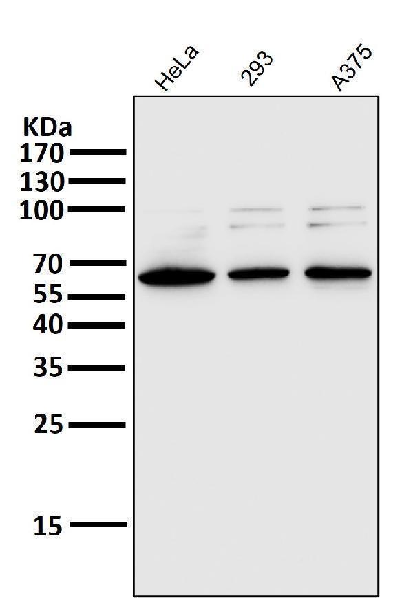
Designing a Western blot for Transcription factor p65? The guide has the expected band size, antibodies backed by real blot images, the controls to run alongside, and protocols taken from published papers.
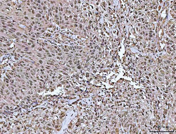
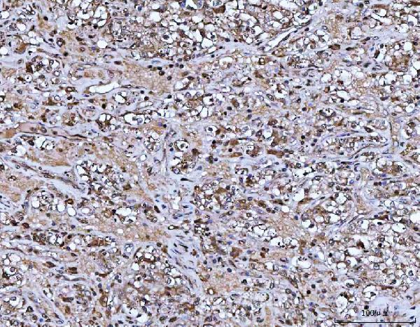
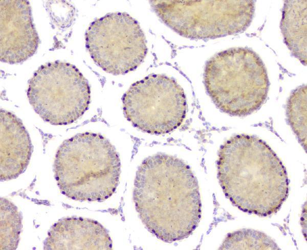
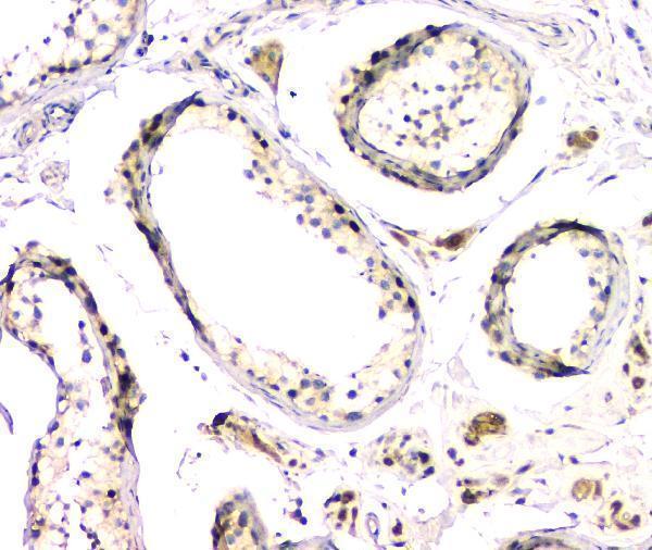
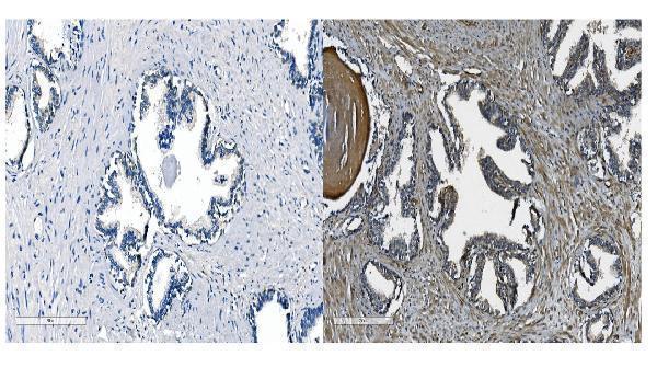
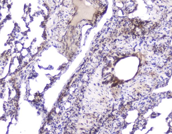
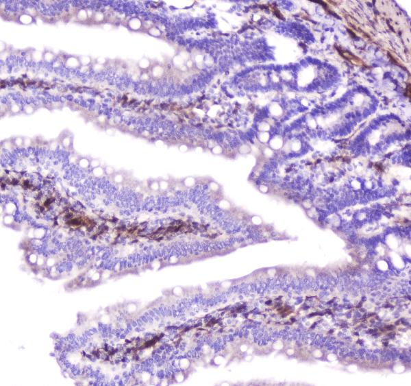
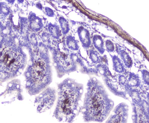
Open the RELA Western Blot GuideDesigning an IHC experiment for Transcription factor p65? The guide has the expected staining pattern, antigen retrieval and fixation, antibodies backed by real IHC images, the tissue controls to run alongside, and protocols taken from published papers.
Open the RELA IHC Guide
95 Citations
38 Citations
21 Citations
140 Citations
18 Citations
42 Citations
23 Citations
26 Citations
26 Citations
10 Citations

10 Citations

8 Citations
8 Citations
7 Citations

12 Citations
5 Citations
5 Citations
5 Citations
7 Citations
8 Citations
6 Citations
9 Citations


6 Citations
3 Citations
3 Citations
3 Citations

13 Citations

6 Citations
6 Citations
2 Citations
2 Citations
4 Citations
5 Citations
5 Citations
2 Citations

2 Citations
2 Citations


3 Citations

4 Citations
4 Citations
8 Citations
4 Citations
1 Citations
2 Citations
3 Citations
3 Citations
3 Citations

3 Citations

3 Citations
1 Citations
2 Citations
3 Citations
1 Citations
1 Citations
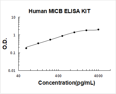
1 Citations

1 Citations
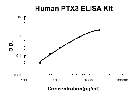
1 Citations
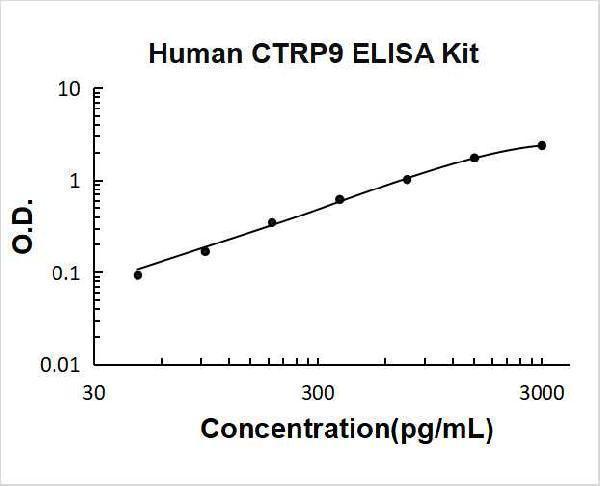
1 Citations
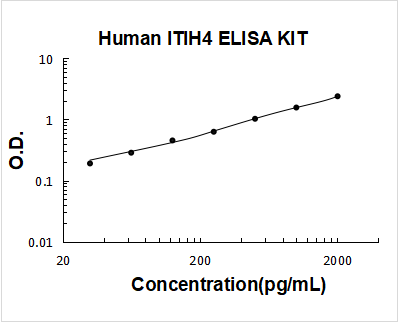
1 Citations
1 Citations
4 Citations
1 Citations
1 Citations
2 Citations
2 Citations
2 Citations
2 Citations

2 Citations
2 Citations
Facts about Transcription factor p65.
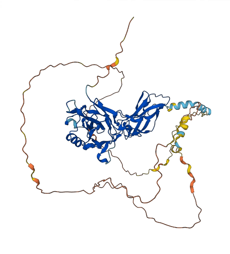
The dimers bind at kappa-B sites in the DNA of their target genes and the individual dimers have distinct preferences for different kappa-B sites which they can bind with distinguishable affinity and specificity. NF-kappa-B is controlled by various mechanisms of post-translational modification and subcellular compartmentalization in addition to by interactions with other cofactors or corepressors.
| Human | |
|---|---|
| Gene Name: | RELA |
| Uniprot: | Q04206 |
| Entrez: | 5970 |
| Belongs to: |
|---|
| No superfamily |

NF kB p65; NFkB p65; NF-kB p65; p65; rela p65; RelA; MGC131774; nf kb p65; NFkB p65; NF-kB p65; NFKB3; NFKB3v-rel avian reticuloendotheliosis viral oncogene homolog A (nuclear factor ofkappa light polypeptide gene enhancer in B-cells 3 (p65)); Nuclear factor NF-kappa-B p65 subunit; Nuclear factor of kappa light polypeptide gene enhancer in B-cells 3transcription factor p65; p65 flow cytometry; p65 immunohistochemistry; p65; p65RelA; rela p65; RelA; v-rel reticuloendotheliosis viral oncogene homolog A (avian); v-rel reticuloendotheliosis viral oncogene homolog A, nuclear factor of kappalight po
Mass (kDA):
60.219 kDA

| Human | |
|---|---|
| Location: | 11q13.1 |
| Sequence: | 11; NC_000011.10 (65653596..65662972, complement) |
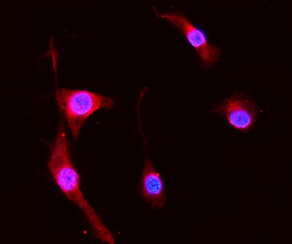
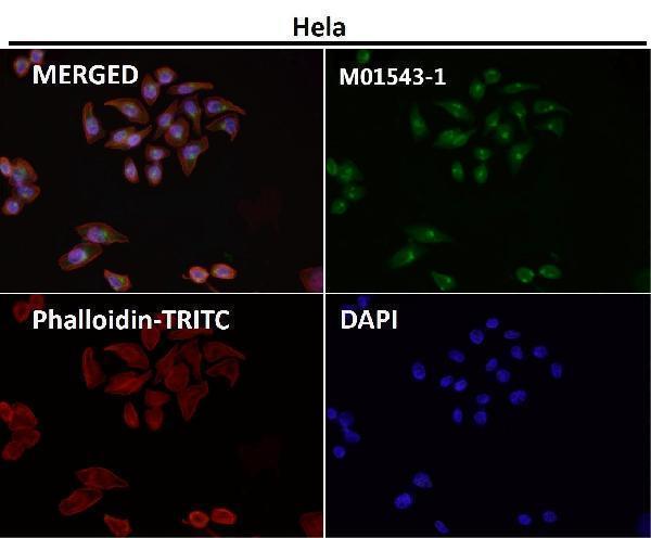
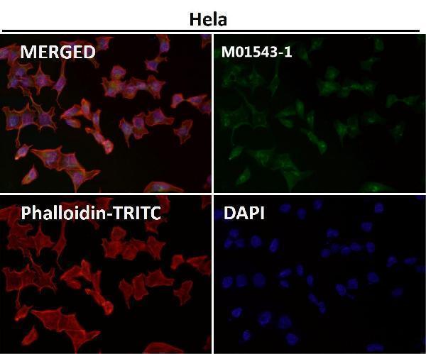
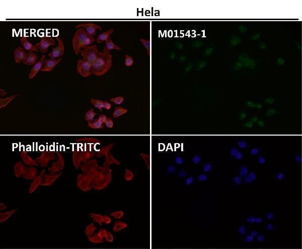
Nucleus. Cytoplasm. Nuclear, but also found in the cytoplasm in an inactive form complexed to an inhibitor (I-kappa-B) (PubMed:1493333). Colocalized with DDX1 in the nucleus upon TNF-alpha induction (PubMed:19058135). Colocalizes with GFI1 in the nucleus after LPS stimulation (PubMed:20547752). Translocation to the nucleus is impaired in L.monocytogenes infection (PubMed:20855622).



PMID: 2006423 by Ruben S.M., et al. Isolation of a rel-related human cDNA that potentially encodes the 65-kD subunit of NF-kappa B.
PMID: 8281153 by Deloukas P., et al. Genomic organization of the gene encoding the p65 subunit of NF-kappa B: multiple variants of the p65 protein may be generated by alternative splicing.
*Showing only the more recent 20. More publications can be found for each product on its corresponding product page